
Hematoma
Hematoma’s appear as anechoic or hypoechoic area overlying the hyperechoic convex line (skull) (figure 8).
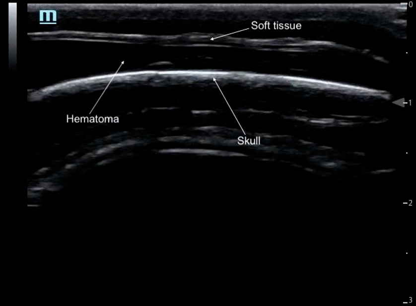 Figure 8: Anechoic hematoma under soft tissue and above hyperechoic convex skull without fracture. Image courtesy of Sick Kids Toronto
Figure 9: Hypoechoic hematoma under soft tissue and above hyperechoic convex skull without fracture.
Skull Fractures
Skull fractures appear as a disruption of the regular smooth convex skull (figure 10). They can appear simple or depressed and often have jagged edges. PoCUS findings regarding fracture type have good correlation to bony window of CT head [1] (figure 13).
· Non-depressed fractures typically present as a linear break without contour change of the skull (figure 11).
· A depressed fracture will appear as a step-off or inward displacement in the normally smooth convex bone surface (figure 12).
Unlike sutures, fractures are not symmetric and should not be found on the contralateral side, they may or may not trace back to sutures/fontanelles. Generally, an associated hematoma is visible in the surrounding soft tissues.
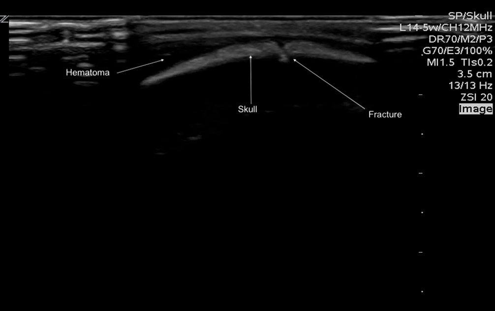
Figure 10: Labeled image of a non-depressed fracture, as seen by a break in the hyperechoic convex skull, with an associated hematoma
Figure 11: Break in the hyperechoic convex skull in a non-depressed fracture with an associated hematoma. Video courtesy of Sick Kids Toronto
Figure 12 Disruption and displacement of the convex hyperechoic skull in a depressed skull fracture with an associated hypoechoic hematoma. Video courtesy of Sick Kids Toronto
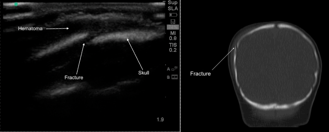
Figure 13: Non-depressed skull fracture with associated hematoma seen on PoCUS (left) and corresponding CT head (right). Note the soft tissue swelling on CT at the fracture site, which corresponds to the hematoma seen on the PoCUS image.