
What is Normal
On ultrasound, the appendix appears as a blind-ended tubular structure in the long axis and a target-like or oval structure in the short axis.
| A normal appendix has the following sonographic features: | |
| – | Thin (≤ 6mm) |
| – | Layered appearance (gut signature). The lumen may contain air, fluid or debris |
| – | Compressible with gentle probe pressure |
| – | Minimal vascularity |
| – | No peristalsis |
| – | Difficult to locate (due to variable tip position) |
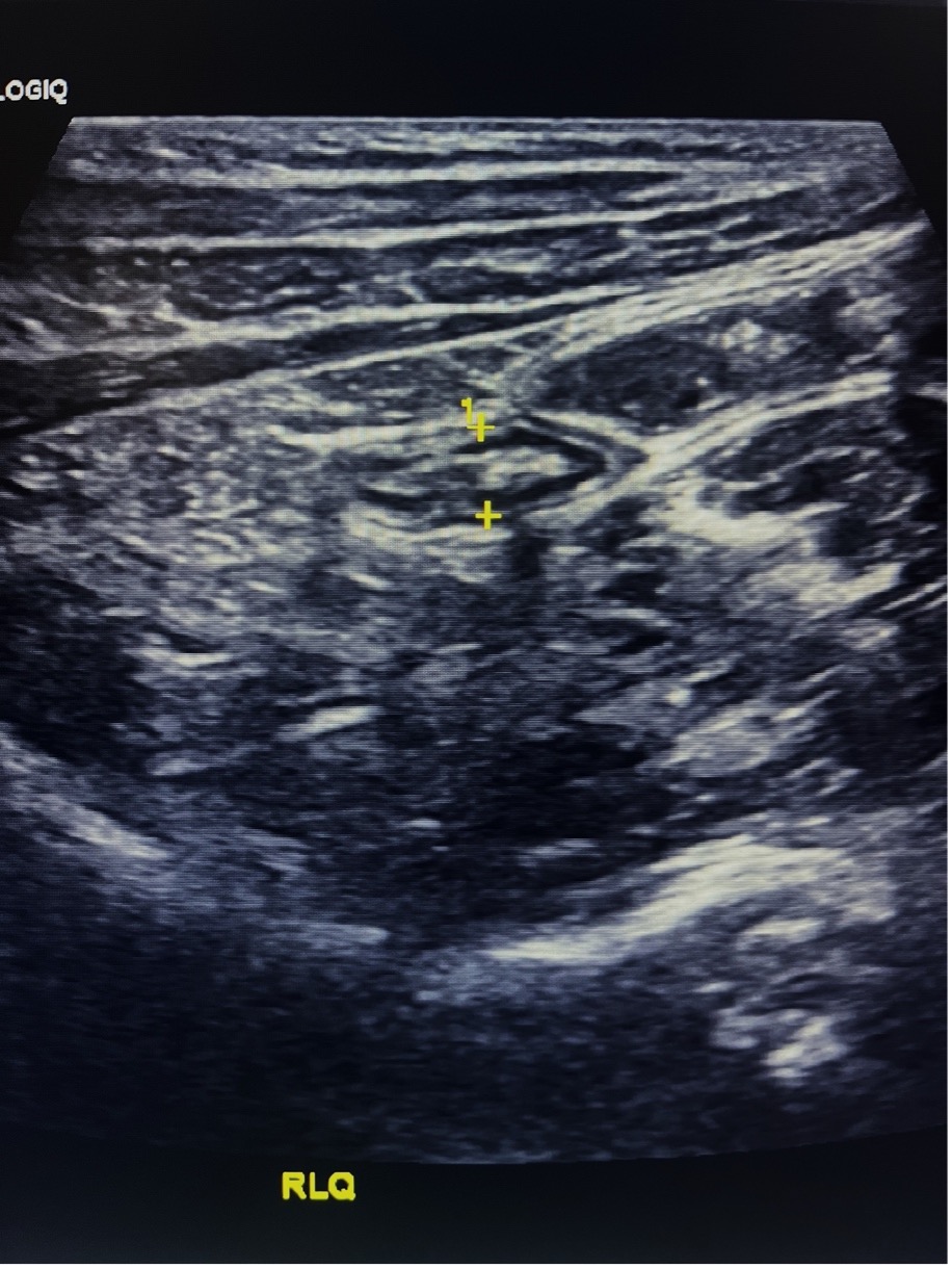
Figure 6. Long-axis view of the appendix tip, demonstrating a normal appearance
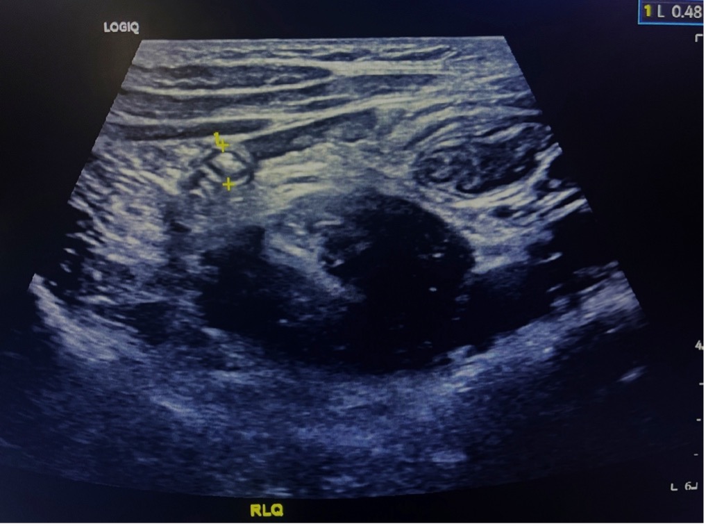
Figure 7 Normal appendix in short axis
Figure 8: Normal appendix in cross-section (yellow arrow) off the colon (pink arrow) medially. Video courtesy of Dave Kirschner, used with permission.