
What is Normal
A normal testicle will be ovoid shaped with a vertical lie. The echogenicity on gray scale US should be uniform and homogonous. You should see the tunica albuginea lining the outside of the testicle as a thin, hyperechoic rim. The mediastinum testis is a hyperechoic band crossing the center of the testicle, best seen in the longitudinal view (Figure 3); however, it may not always be visible, and in the setting of trauma, it can be mistaken for pathology. The epididymis is seen adjacent to the testis and is slightly hypoechoic relative to the testis (figure 4). In color flow doppler, the testicles should have blood flow equal bilaterally. Look for presence of arterial blood flow in the center of the testicle (Figure 5). Peripheral capsular vessels are often larger and easier to detect on Doppler imaging than the smaller intratesticular vessels. Symmetric flow should be identified within the central testicular parenchyma, not just around the periphery.
In prepubertal children, intratesticular blood flow is often low-flow and more difficult to detect, even in healthy testicles. Smaller vessel size and slower flow velocities can make Doppler assessment technically challenging, particularly with PoCUS or lower-sensitivity ultrasound systems. Careful optimization of Doppler settings and direct side-to-side comparison are especially important in this age group. If similarly low-flow appearances are seen bilaterally, this may reflect normal prepubertal physiology. In contrast, low flow on only one side should raise concern for pathology, including torsion.
If blood flow is difficult to identify or findings are uncertain, store representative clips and images and proceed with formal diagnostic ultrasound assessment.
The spermatic cord should appear as a smooth linear echogenic structure in the longitudinal plane and as multiple small round or oval tubular structures in the transverse plane. On color Doppler imaging, vascular flow should be visible within the spermatic cord vessels.
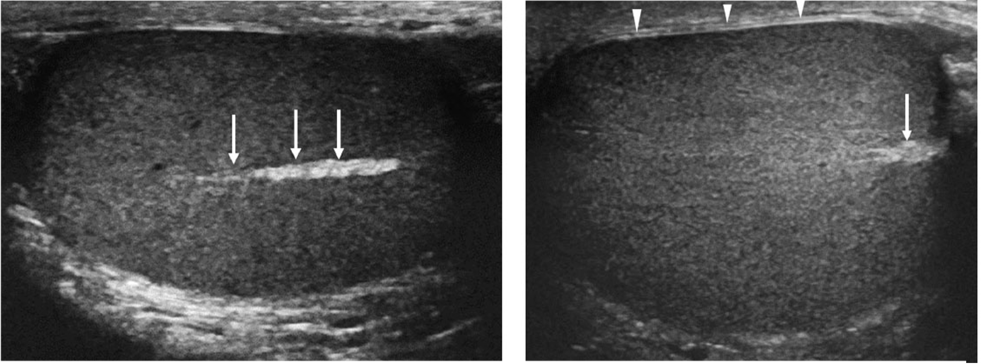
Figure 3. Normal testicles in longitudinal view. The arrows are pointing to the mediastinum testis which is a normal finding. The arrowheads are outlining the tunica albuginea [13]
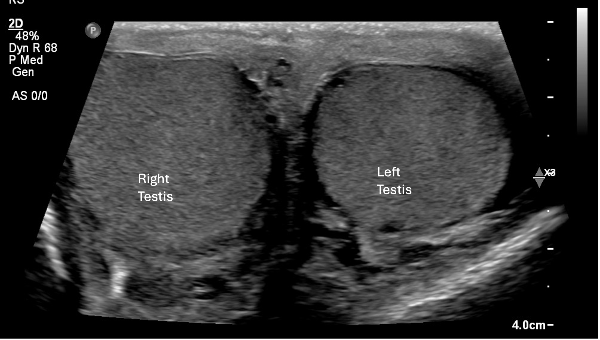
Figure 4. Normal testicles in transverse view
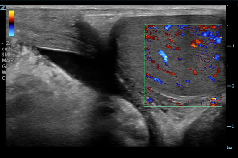
Figure 5. Color flow doppler of normal testicle in longitudinal view
Video 1: Normal color doppler of the left testicle. Video courtesy of David Kirschner, used with permission.