
In both planes, posterior structures appear in the near field (top of the image) and anterior structures in the far field (bottom of the image). In the sagittal orientation, screen left corresponds to the cephalad and screen right to caudal. In the transverse orientation, screen left corresponds to the patient’s left, and screen right to the patient’s right.
The appearance of the spinal anatomy on ultrasound differs significantly between neonates/infants and older children. These differences are mainly due to the degree of ossification, visibility of ligamentous structures, and the relative brightness of bony elements.
Neonates & Infants
Sagittal View
· Spinous processes – Poorly ossified in this age group, appearing assmall, rounded, low-echogenicity structures with little to no acoustic shadowing.
· Interspinous space – Typicallywide and clearly visualized hypoechoic gaps between the under-ossified spinous processes. This is the window through which the needle passes.
· Dura – a thin, echogenic linear structure forming the posterior border of the subarachnoid space
· Subarachnoid (CSF) space – awell-defined anechoic fluid filled space posteriorly in the spine
> Contains the Cauda equina nerve roots
· Cauda equina – mobile, linear hyperechoic strands within the subarachnoid space/CSF
> Cauda equina pulsationsmay be visible within the dura
· Conus medullaris – The tapering end of the spinal cord, appearing as a hypoechoic cord-like structure that transitions into the cauda equina.
> In neonates/infants it typically lies more cranially (L1–L2), but can occasionally be slightly lower.
· Filum – fine echogenic line extending inferiorly from the tip of the conus within the hypoechoic CSF.
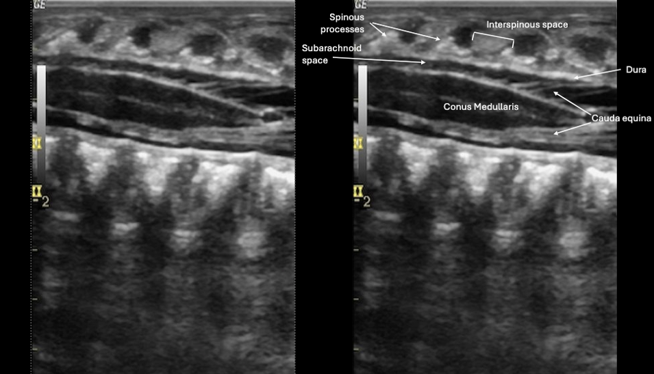 Figure 5. Sagittal view of the neonatal spine on PoCUS. Image adapted from Radiopaedia, courtesy of Kewal Arunkumar Mistry, Radiopaedia.org, rID: 34886. Used under license CC-NC-BY-SA 3.0
Transverse View
· Spinous process – Appears as a small, rounded hypoechoic structure in midline of the near field. It will have minimal or absent posterior shadowing.
· Transverse processes – Bilateral, lateral mildly echogenic lines producing some acoustic shadowing.
> Less prominent than in older children due to incomplete ossification.
· Dura – a thin, echogenic linear structure forming the posterior border of the subarachnoid space
· Subarachnoid Space – well-defined anechoic fluid filled space posteriorly in the spine
> Contains the Cauda equina nerve roots
· Conus medullaris – Hypoechoic, round central structure within the subarachnoid CSF space
· Cauda equina – small, echogenic dot-like structuresdistributed around the central CSF space, surrounding the conus medullaris
> Typically mobile, showing subtle movement with CSF pulsations.
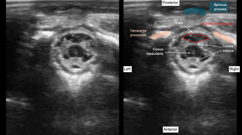 Figure 6. Transverse view of the neonatal spine on PoCUS. Image adapted from Radiopaedia, courtesy of Kewal Arunkumar Mistry, Radiopaedia.org, rID: 34886. Used under license CC-NC-BY-SA 3.0
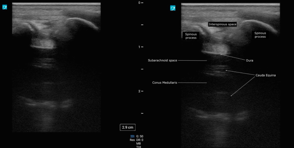 Figure 7. Sagittal view of the spine of a 6-month old infant. Note the reduced visualization of spinal anatomy compared with the neonatal spine.
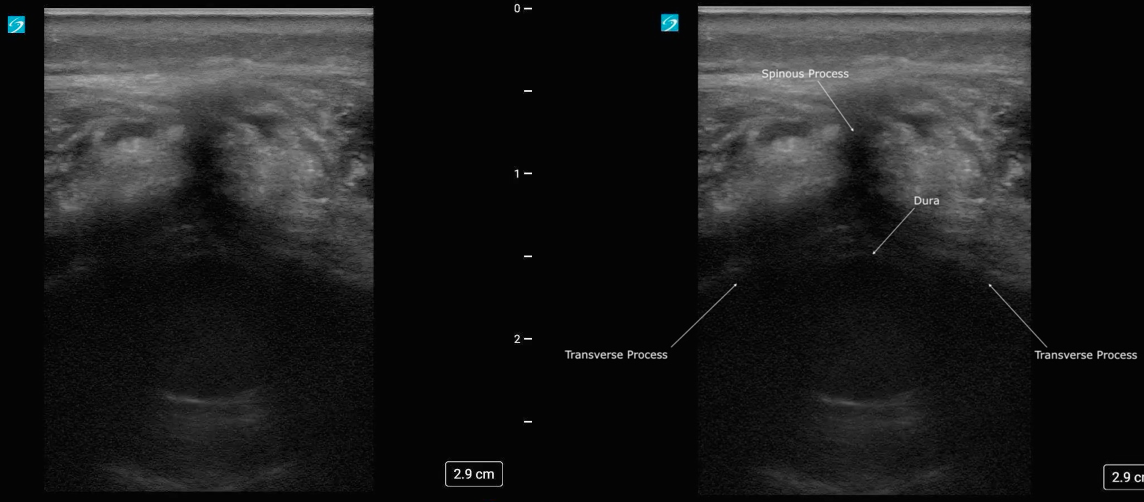 Figure 8. Transverse view of the spine of a 6-month old infant. Note the reduced visualization of spinal anatomy compared with the neonatal spine.
Children
Sagittal View
· Spinous processes – curved, bright echogenic lines deep to the subcutaneous tissue that cast posterior acoustic shadows
· Interspinous space – hypoechoic gaps between adjacent bright spinous process shadows. This is the window through which the needle passes.
· Ligamentum flavum – visualized as a single, distinct echogenic band deep to the interspinous space. It is not always visible, but when seen, it represents the target depth for needle insertion.
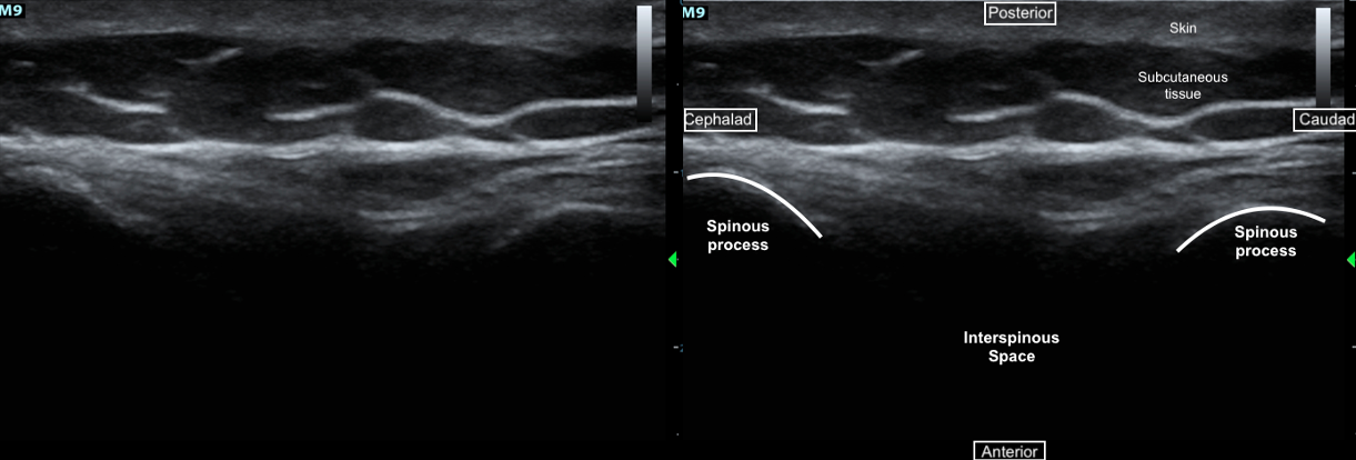 Figure 9. Sagittal view of the spinous processes.
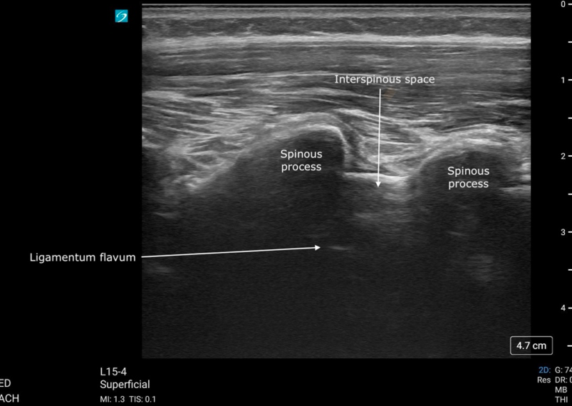
Figure 10. Sagittal view of the spinous processes and ligamentum flavum
Figure 11. Sagittal clip of the spine
Transverse View
· Spinous processes – a hyperechoic central structure in the near field with dense acoustic shadowing beneath, marking the midline.
· Laminae – paired echogenic arcs lateral to the spinous process
*Note, in younger children, the laminae are oriented more obliquely toward the midline, so they appear as continuous oblique hyperechoic lines that run diagonally toward the spinous process (figure 13).
· Transverse processes – more lateral echogenic lines with posterior shadowing, seen at greater depth.
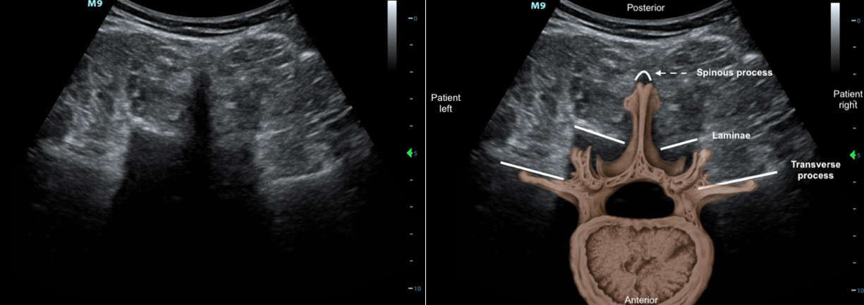
Figure 12. Transverse view of the vertebrae including the spinous process, laminae and transverse process
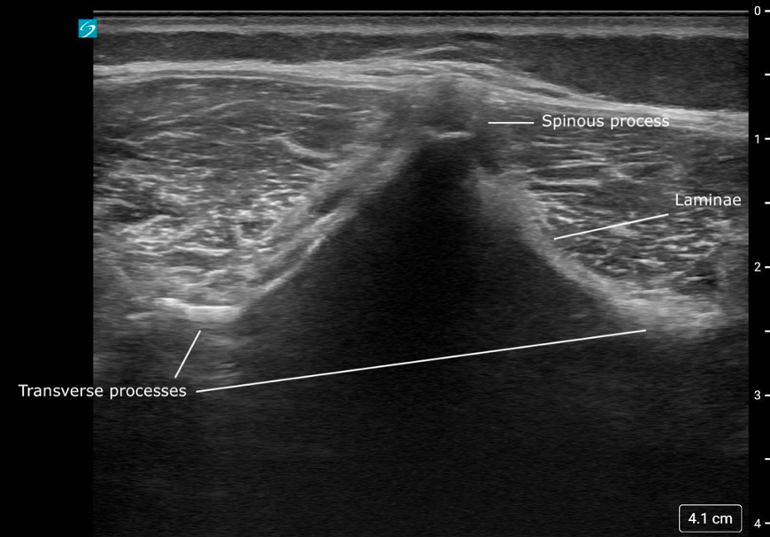
Figure 13. Transverse view of the vertebrae in a younger child, with more oblique appearing laminae
Figure 14. Transverse sweep of the spine in a young child, labeled
Figure 15. Transverse slide along the lumbar spine, with each vertebra coming into view as the probe moves caudally.

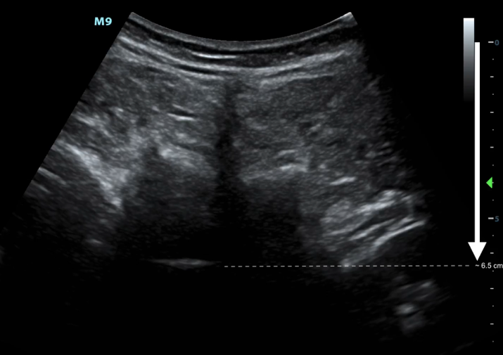 Figure 16. Transverse view of the vertebrae including the ligamentum flavum with the depth scale on the right side of the screen used to estimate the required needle insertion depth as roughly 6.5 cm.