
Hip Anatomy Review
The hip joint is a ball and socket joint, comprised of the femoral head and neck and the acetabulum (made up of the ischium, pubis and ilium). The joint capsule surrounds these structures and contains synovial fluid (Figure 5).
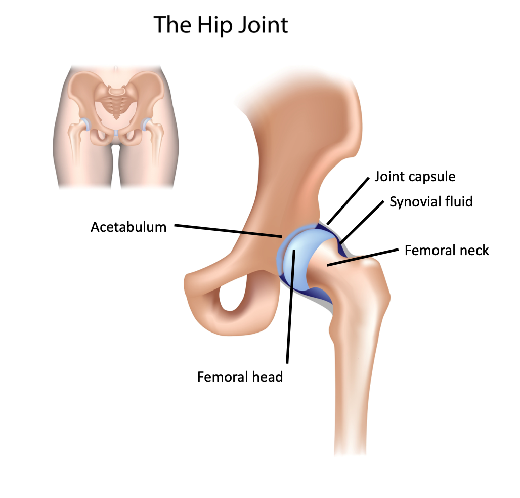 Figure 5: Anatomy of the hip joint
Superficial to the bony structures, we find the musculature of the pelvis and the hip. Those that are relevant to us for the purpose of scanning for hip effusions include the iliopsoas, quadriceps and sartorius. These muscles are found superficial to the joint (Figure 6).
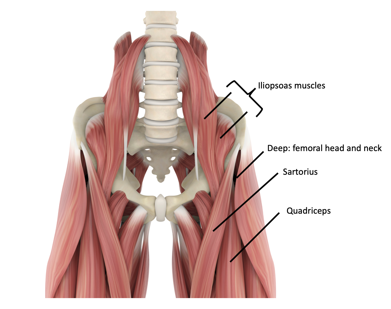 Figure 6: Hip musculature
Technique
- Position the patient supine, with the affected hip in a neutral position [4].
- Place the linear array probe below the femoral crease, with the probe at a 45-degree angle and pointing posteriorly on the patient: parallel to the neck of the femur (Figure 7).
- Identify the femoral neck, femoral head, iliopsoas and synovial space in between [7].
- Assess for presence of joint effusion.
- Repeat on the contralateral joint.
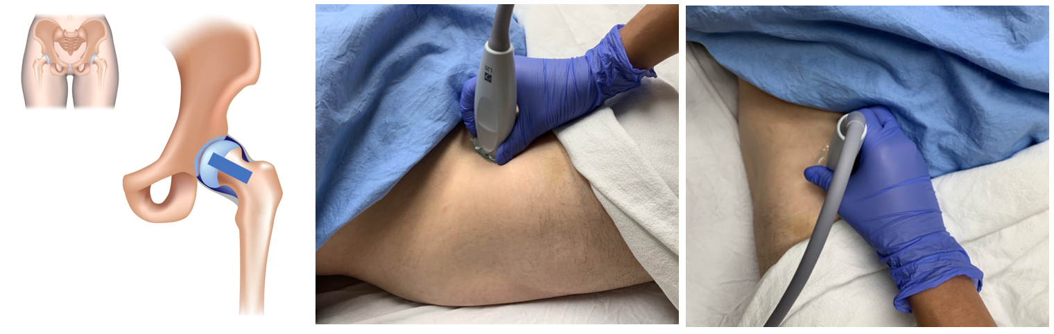
Figure 7: Probe position for hip ultrasound
What am I looking at?
Femoral neck
- Hyperechoic horizontal line.
Femoral head
- Curved hyperechoic line.
- Physis or growth plate can be seen as a notch or hypoechoic break in the femoral head [7].
Muscles
- Iliopsoas muscle is superficial to the synovial space.
- Quadriceps and sartorius are the most superficial structures in the field of view.
Synovial space
- Space between the femoral neck and the iliopsoas muscle.
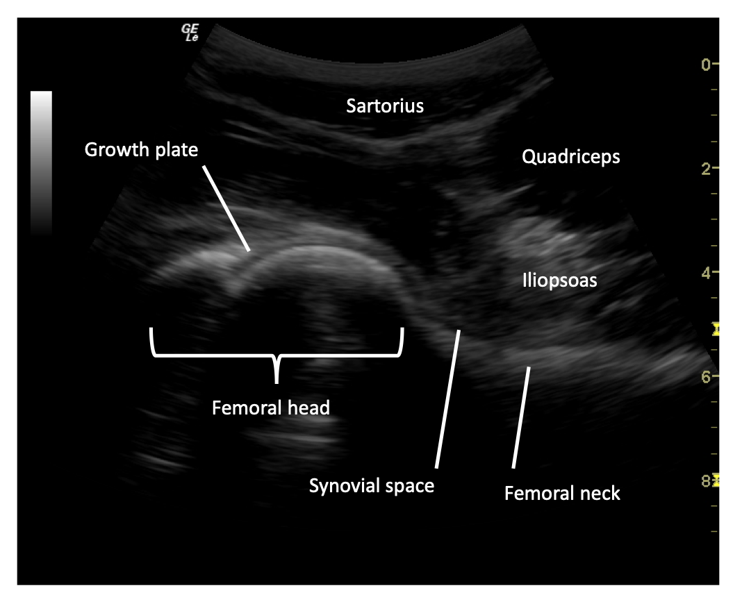 Figure 8: Labelled normal hip ultrasound.
What is normal?
Normally, synovial fluid will follow the contours of the joint itself, in this case, the femoral head and neck. The size of a fluid collection in the hip is measured from the anterior surface of the femoral neck to the posterior surface of the iliopsoas muscle [4]. The space should be <5mm and within 2mm in comparison to the unaffected hip (Figure 9).
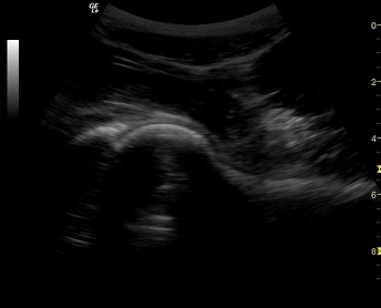 Figure 9: Normal adult hip ultrasound.
 Figure 10. Normal pediatric hip ultrasound appearances in children aged 2 and 4 years, demonstrating age-related differences in the amount and appearance of cartilage and ossification. The examples illustrate the progressive ossification of the developing hip, with relatively greater hypoechoic cartilage in the younger child and increasing echogenic ossification with age.
What is NOT normal?
The presence of a joint effusion in a hip is defined as a space between the anterior surface of the femoral neck to the posterior surface of the iliopsoas muscle of >5mm or >2mm difference in comparison to the unaffected side (Figure 11). An effusion will classically have a convex appearance (Video 2) in comparison to the normal concave appearance of the synovial fluid in a joint without an effusion.
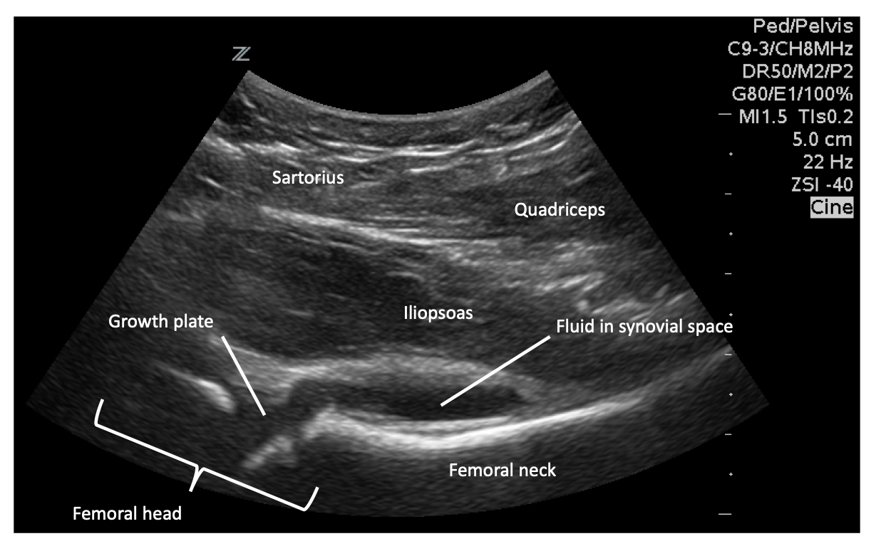 Figure 11: Positive hip effusion.
Video 1: Hip joint effusion. Note the convex appearance of the fluid on the image on the right.