
Normal skin and soft tissue have many layers (Figure 1). The most superficial structures are the epidermis and dermis, which appear as if one hyperechoic structure. Deep to this, you will find subcutaneous fat, which is hypoechoic and globular. Blood vessels, nerves and lymph nodes are found within the hypodermis, and are varying in their echogenicity. Deep to these structures, the muscle is found beneath a hyperechoic layer of fascia. Muscle is seen as highly organized hypoechoic and striated fibers.
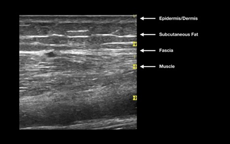 Figure 1: Anatomy of normal skin
Tendons are visualized as hypoechoic fibrillar organized structures, whereas fat pads are also hypoechoic, but are more homogenous in their appearance (Figure 2). Finally, smooth hyperechoic bone cortex may be seen as the deepest layer.
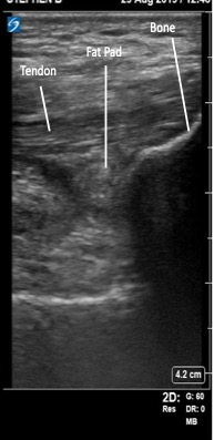 Figure 2: Anatomy of a normal joint
Special Image Characteristics in Pediatric MSK Ultrasound
Children’s joints contain the same basic components as adult joints, but their appearance differs because the epiphyses in children are partially or completely composed of hyaline cartilage and growth plates remain open until the end of puberty [9].
In young children, the cartilaginous epiphysis is thick, and secondary ossification centres are small, giving the epiphysis a predominantly anechoic-to-hypoechoic appearance [9,10]. These secondary ossification centers gradually enlarge through adolescence, creating mixed echogenicity before the epiphysis becomes fully ossified in adulthood [9]. Because of the hypoechoic appearance and the increased cartilage thickness in early childhood, the expected depth to the joint capsule and can mimic or obscure joint effusions if not recognized [10]. The cartilaginous epiphysis may also have visible blood vessels on color doppler in young children [9].
Growth plates, which appear as hypoechoic ‘breaks’ or notches between the metaphysis and epiphysis, may also show irregularities in older children that should not be mistaken for the joint space [10].
Key point: Age-dependent differences in cartilage thickness, ossification patterns, and growth plate appearance are normal features of pediatric MSK ultrasound. Recognizing these features helps prevent normal cartilage or physeal anatomy from being misinterpreted as joint fluid or other pathology.
 Figure 3ab. A) Normal pediatric (2 yrs) non-ossified knee VS (B) normal adult knee
Video 1. Normal pediatric knee ultrasound in a 4-year-old demonstrating hypoechoic cartilaginous portions of the distal femoral epiphysis and patella, with an echogenic patellar ossification center and adjacent hypoechoic developing ossification.
Scanning Tip: Scan with dual screen functionality
In order to facilitate comparing an affected joint with the contralateral side, it can be useful to use the dual screen function on the ultrasound machine.
- Choose “More Controls” from the main screen, found at the bottom right corner (Figure 4a).
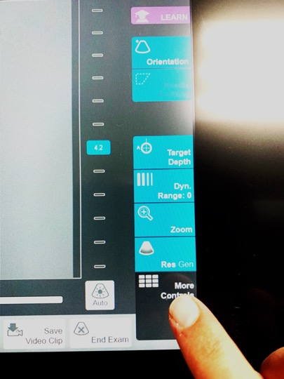 Figure 4a: Choose “more controls”
 - Select “Dual” from the options screen (Figure 3b)
 Figure 4b: Choose “dual”
 - Touch the screen on either side to start scanning. When ready to switch, freeze the area of interest on the screen and then touch the opposite screen to begin scanning the opposite side (Figure 4c).
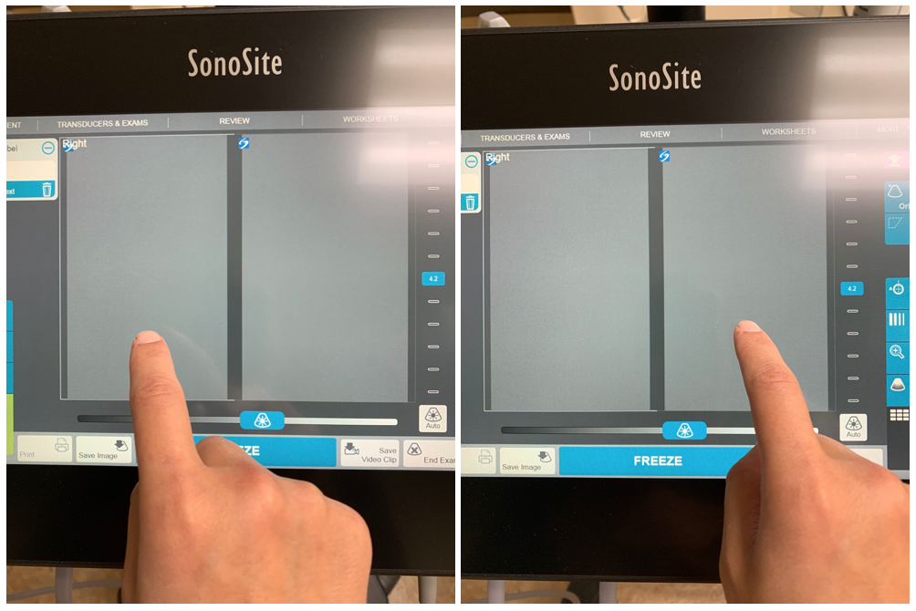 Figure 4c: Toggle between screens by touching the desired side
 - Below is an example of a split screen view (Figure 4d).
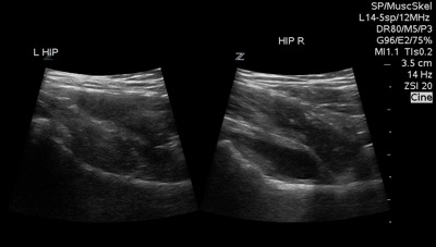 Figure 4d: Dual screen example