
Begin with Unaffected Testicle
1. Apply a COPIOUS amount of gel over the scrotum
2. 2D Grey scale assessment: Start in 2D gray scale mode and scan each testicle in both the transverse and longitudinal planes (Figure 2). This can be used to assess testicle size, lie and texture.
· Transverse orientation: probe marker pointed to patient RIGHT.
· Longitudinal orientation: probe marker pointed CRANIALLY.
3. Evaluate with color Doppler: In the longitudinal view, select the color doppler function to assess flow to the testicle.
· Be sure to use a low flow Doppler setting and adjust the color gain as needed to optimize visualization—ensure it’s neither too low, which may cause missed flow signals, nor too high, which can obscure the image with noise.
4. Evaluate with spectral Doppler (if required by your local policy): with the color Doppler still activated, select the pulsed-wave Doppler function and place the sample box over a prominent vessel. Record a spectral tracing of arterial and venous flow (if possible).
5. Evaluate the Spermatic Cord: In the longitudinal plane, slide the probe superiorly along the length of the spermatic cord. Use greyscale and color Doppler to assess for abnormal twisting.
· Store clips in both planes regardless of whether it appears normal or abnormal.
6. Buddy View or Dual Testicle Comparison: If you have a large linear probe, position the probe in the transverse plane at the inferior scrotum and apply minimal upward pressure to lift both testicles. This allows for direct visualization of both testicles simultaneously to compare size, texture, lie, and color Doppler flow.
· If using the small linear probe, assess each testicle individually and save images to compare—be careful to not change settings on the machine between sides.
· Compare to the affected testicle for increased, decreased, or absent flow
· Obtain both 2D and color Doppler clip or image
Repeat steps 2-5 on the unaffected testicle
Be sure to document and store images at each step of the exam, including both the affected and unaffected sides
Scanning Tips:
- Optimize color Doppler settings (including gain and PRF/scale) on the unaffected testicle first to achieve the best possible visualization of normal intratesticular flow prior to comparing sides.
- Be sure NOT to change any of the color Doppler settings between the unaffected and affected testicles, as changes in gain, scale, or other Doppler parameters can create the false appearance of differences in vascularity and make side-to-side comparison unreliable.
- Be mindful that capsular vessels are often larger and easier to detect on Doppler imaging than intratesticular vessels. Ensure color Doppler demonstrates flow within the central testicular parenchyma, and that spectral Doppler sampling is obtained from an intratesticular vessel rather than a capsular vessel, as capsular flow may still be preserved in early, partial or intermittent torsion.
- Venous flow may be difficult to capture on spectral Doppler. Make up to three attempts; if venous flow cannot be obtained, proceed with the examination
- It is best practice to clearly label all images and clips as left or right at the time of acquisition to ensure accurate interpretation and side-to-side comparison during review by radiology and urology.
- Use a very light probe pressure to avoid compressing, displacing or rotating the testicle. You can rest your elbow or forearm on the patient’s legs or a stack of towels to help stabilize your hand and maintain gentle, controlled pressure.
- The testicles are mobile structures, so their position and orientation within the scrotum may vary between sides. When obtaining a buddy view, the probe may need to be rotated slightly clockwise or counterclockwise to accommodate their position and ensure both testicles are within the field of view
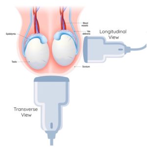
Figure 2. Ultrasound image acquisition of the testicles in the transverse and longitudinal planes. Note, the probe positions shown are a general guide and may be adjusted based on the position of the testicle. Sagittal views may be obtained from either the lateral or anterior scrotum, while transverse views may be obtained from either an inferior or anterior approach.