
Ankle Anatomy Review
The area where an effusion in the ankle collects is at the intersection of the tibia and the talus (Figure 18). There is a fat pad (the anterior fat pad) at the junction of these two bones. Superficial to the boney surfaces, the tibialis anterior tendon is found.
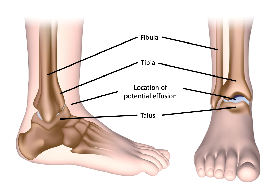 Figure 18: Anatomy of the ankle joint.
Technique
- Position the patient supine.
- Have the patient flex their knee and place their foot on the bed to place their ankle in dorsiflexion [1].
- Place the linear array probe along the longitudinal axis of the tibia at the joint line between the tibia and talus [1] (Figure 19).
- Identify synovial space at the junction of the talus and the tibia.
- Assess for presence of joint effusion.
- Repeat on the contralateral joint.
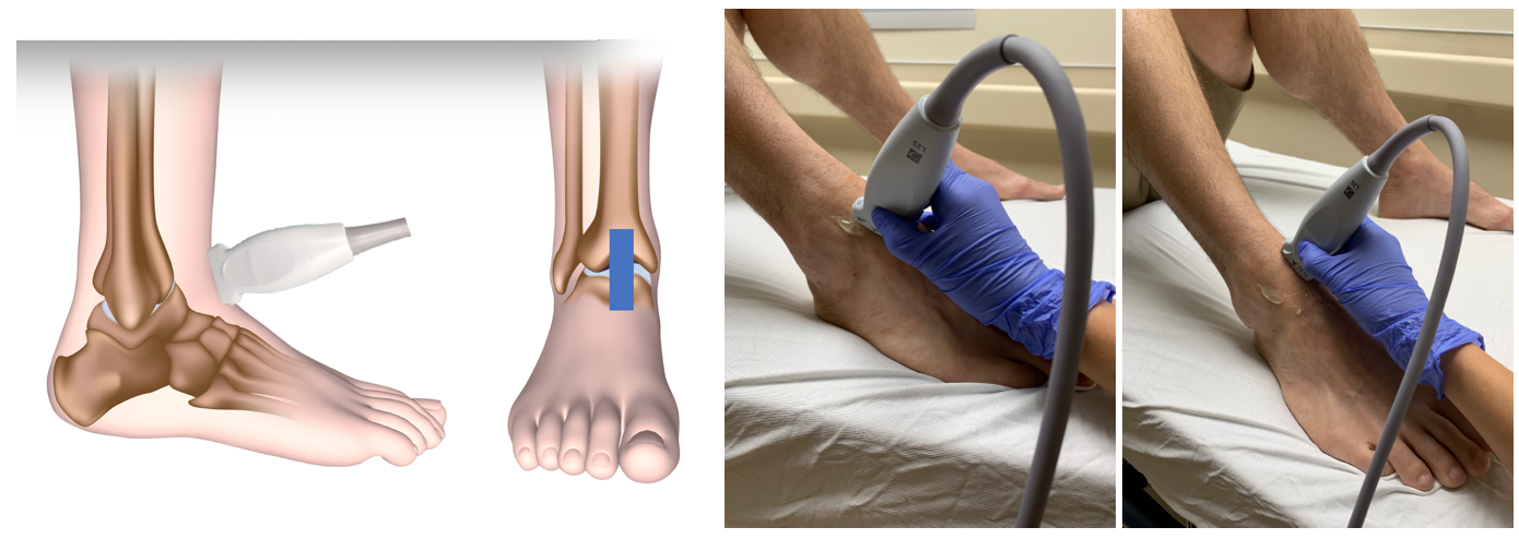 Figure 19: Probe position for ankle ultrasound.
What am I looking at?
Tibia
- Hyperechoic line cephalad.
Talus
- Hyperechoic line caudal.
Tibialis anterior tendon
- Fibrillar hyperechoic structure in near field.
Anterior fat pad
- Immediately superficial to the synovial space.
Anterior talotibial recess or synovial space
- Superficial to the bony surfaces of the joint.
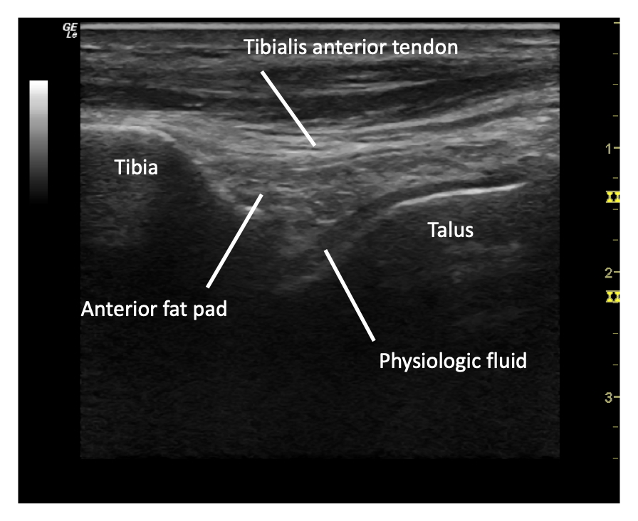 Figure 20: Labelled normal ankle ultrasound. Note that physiologic fluid and cartilage can appear similarly, so differentiate them with compression of the structures. Fluid will compress, cartilage will not.
What is normal?
On ultrasound of the ankle, you will see the tibia extending cephalad and the joint line between the tibia and the talus (Video 5). Within the joint space, near field to the bony structures, there may be a small amount of physiologic fluid, and an anterior fat pad. As in the other joints, physiologic fluid will follow the contours of the bone, and have a more concave appearance (Figure 21).
Video 4: Normal ankle ultrasound.
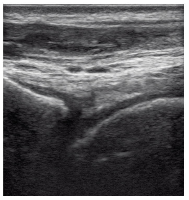 Figure 21: Normal ankle ultrasound.
 Figure 22. Normal pediatric ankle ultrasound appearances in children aged 2, 4, and 8 years, demonstrating age-related changes in the amount and appearance of cartilage and ossification. The examples illustrate the progressive ossification of the developing skeleton, with relatively greater hypoechoic cartilage in younger children and increasing echogenic ossification with age
What is NOT normal?
An ankle effusion is characterized by a convex shaped hypoechoic fluid collection anterior to the tibial and talar joint (Figure 23). In the ankle, unlike in the hip, there is no specific measurement for a positive effusion – the important feature to look for is the convexity of the collection (Video 6).
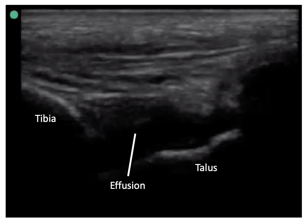 Figure 23: Positive ankle effusion.
Video 6: Ankle joint effusion. Again, note the convex appearance of the fluid.