The information presented in this section is based primarily on Health Canada’s Guidelines for the Safe Use of Diagnostic Ultrasound (2001) [10]. Where applicable, additional sources have been cited to supplement or update specific recommendations.
ALARA
ALARA stands for As Low As Reasonably Achievable and is the guiding principle for safe ultrasound practice. The goal is to obtain the diagnostic information needed while minimizing unnecessary exposure. In diagnostic ultrasound, ALARA refers to using the lowest combination of acoustic output and exposure time necessary to answer the clinical question [10].
Foundations of Ultrasound Safety
Diagnostic ultrasound has an excellent safety record and does not use ionizing radiation. However, ultrasound transfers energy into tissues and, under certain conditions, may produce biological effects. Understanding these effects helps clinicians use ultrasound safely.
Ultrasound energy can interact with tissue through two primary mechanisms: thermal effects and mechanical effects.
1) Thermal Effects
Thermal effects occur when tissues absorb ultrasound energy and convert it to heat. The amount of heating depends on both the ultrasound exposure and the type of tissue being scanned.
Bone absorbs ultrasound energy more readily than soft tissue, while fluids absorb very little. As a result, temperature increases are of greater concern when bone is present within the ultrasound beam, including fetal bone during pregnancy. Because bone absorbs ultrasound energy efficiently, less acoustic power is required to produce a temperature rise than in soft tissue [11].
Heat may also be generated by the transducer itself. Additional caution may be warranted during transvaginal, transrectal, and transesophageal examinations, as transducer heating may contribute to local tissue warming. Similar considerations apply when bone is located close (within 1cm) to the transducer surface, such as during ocular or cranial imaging.
2) Mechanical Effects
Mechanical effects are non-thermal interactions caused by pressure changes within the ultrasound wave. The primary concern is cavitation, which refers to the response of gas bubbles exposed to alternating pressure cycles. Cavitation may occur as stable cavitation, where bubbles repeatedly expand and contract, or inertial cavitation, where bubbles grow and collapse rapidly [11].
In routine diagnostic ultrasound, cavitation is considered unlikely in most tissues because naturally occurring gas bubbles are generally absent. The theoretical risk is greater in tissues containing gas, such as the lungs or bowel, and when ultrasound contrast agents containing microbubbles are used.
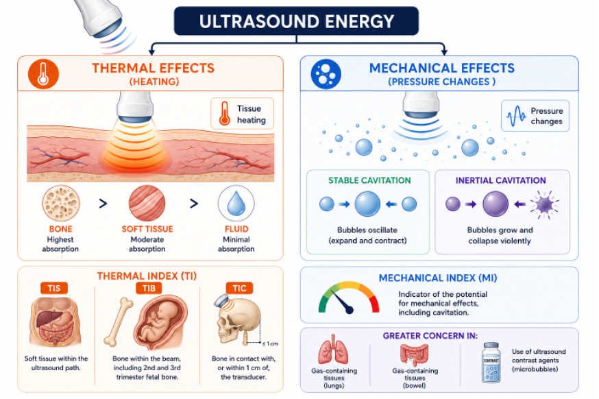 Figure 1. Ultrasound safety basics
Machine Parameters
Several parameters influence ultrasound exposure and are displayed on modern ultrasound systems.
Dwell Time
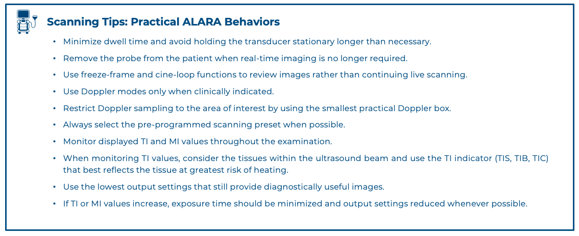
Dwell time refers to the amount of time the transducer actively transmits ultrasound while remaining over a particular area. Dwell time is one of the most important factors affecting tissue exposure and can be readily controlled by the operator.
Thermal Index
The Thermal Index (TI) is an indicator of the potential for tissue heating. It represents the ratio of the emitted ultrasonic power to the power required to raise tissue temperature by approximately 1°C under specific model conditions.
The TI indicator is displayed on the ultrasound screen.
Different TI’s are used depending on the tissue being examined:
· TIS (Soft Tissue):used when primarily soft tissue is within the ultrasound path.
· TIB (Bone):used when bone, including 2nd and 3rd trimester fetal bone, is within the beam.
· TIC (Cranial):used when bone is in contact or within 1cm of the transducer.
Because bone absorbs ultrasound energy more readily than soft tissue, TIB and TIC are particularly important when scanning fetal, neonatal, or cranial structures.
Mechanical Index
The Mechanical Index (MI) is an indicator related to the potential for mechanical effects, including cavitation. It reflects the relationship between ultrasound pressure and transducer frequency.
Modern diagnostic ultrasound systems are designed with a maximum attainable MI of 1.9, a limit intended to substantially reduce the likelihood of clinically significant mechanical injury.
The MI indicator is displayed on the ultrasound screen
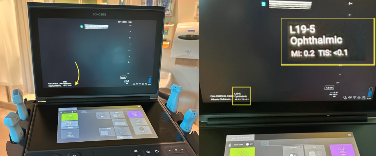 Figure 2. MI and TIS as seen on a SonositePX PoCUS unit.
Output Power
Output power refers to the amount of ultrasound energy emitted by the transducer, or acoustic output. Increasing output power increases the amount of energy delivered to tissues and may increase the potential for thermal or mechanical effects.
Scanning tip: Gain vs Output Power
Gain and output power are not the same. Gain adjusts how returning echoes are displayed on the screen and affects image brightness without increasing patient exposure.
Output power changes the amount of ultrasound energy transmitted into the patient. Increasing the power directly on the machine is not recommended.
B-Mode and Doppler Modes
Different imaging modes produce different levels of acoustic output.
· B-mode generally produces the lowest acoustic output.
· M-mode may produce higher outputs than B-mode.
· Color Doppler and pulsed-wave Doppler typically generate higher acoustic outputs and have a greater potential for tissue heating.
Although M-mode and Doppler are valuable clinical tools, their use should be guided by the ALARA principle, particularly when imaging sensitive tissues [12].
Pulse Repetition Frequency (PRF):
PRF refers to the number of ultrasound pulses transmitted by the transducer each second. PRF is most commonly adjusted during Doppler examinations and contributes to the higher acoustic outputs associated with Doppler imaging. Increasing PRF increases the number of ultrasound pulses transmitted per second and may increase acoustic exposure.
Scanning tip: On many ultrasound systems, the Doppler scale control is the user-facing adjustment for PRF. Increasing the scale typically increases the PRF, allowing higher velocities to be displayed and reducing aliasing, while decreasing the scale lowers the PRF and improves sensitivity to slower flow.
Frequency
Lower-frequency transducers provide greater penetration but may increase TI and MI because more acoustic energy is required to image deeper structures. Use the highest frequency that adequately answers the clinical question.
Focus Position
The focal zone is the region where the ultrasound beam is narrowest and most concentrated. Moving the focus deeper requires the system to concentrate energy at greater depths, which may increase TI and MI [13]. Position the focal zone at or just below the structure of interest whenever possible.
Scan Area
Scan area refers to the width or size of the imaging field. Reducing the scan area concentrates ultrasound energy into a smaller region and may increase TI and MI. , particularly when the zoom box is narrow and deep [13].
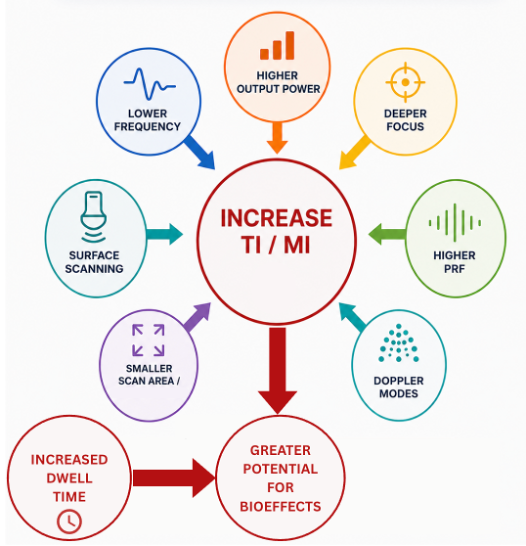 Figure 3. Parameters affecting TI/MI and overall theoretical risk of ultrasound
Special Safety Considerations
Ocular Ultrasound
The eye is particularly sensitive to thermal effects because the lens and intraocular fluids have limited cooling blood supply [12]. Because of this, it is essential to maintain a very low MI and TIS and avoid Doppler during ocular scanning to minimize the theoretical risk of tissue damage [11]. These precautions are particularly important in pediatric ocular examinations.
Neonatal and Pediatric Imaging
Additional caution is recommended when performing ultrasound examinations in neonates, infants and young children. Certain tissues and organs may be more susceptible to potential biological effects of ultrasound, either because they are still developing (such as the brain and spinal cord) or because they contain gas (such as the lungs or bowel) [12]. These considerations that are particularly relevant given the frequent use of cranial, lung, and bowel ultrasound in neonatal and pediatric practice.
When imaging pediatric patients, examinations should be clinically justified, performed efficiently, and exposure times kept as short as reasonably achievable.
Brain, head, and spine:
There is a small risk of thermal effects on the developing neonatal brain, head and spine if the TIC exceeds 0.7 [12].
Lungs:
There is a small potential risk of capillary hemorrhage during ultrasound examinations involving the neonatal or infant chest if the MI exceeds 1.0 [10].
Bowel
Ultrasonically induced capillary hemorrhaging of the intestine has been associated with conditions that inhibit intestinal peristalsis. Clinically significant intestinal hemorrhage is unlikely at diagnostic exposure levels (MI ≤ 1.9), but risk may increase with impaired motility or increased intraluminal gas [10].
Pregnancy
Ultrasound is widely used during pregnancy and has an excellent safety record. However, additional caution is recommended because the developing embryo and fetus may be more susceptible to thermal effects.
For specific guidelines, please refer to Health Canada guidelines [10].
Ultrasound Contrast Agents
Ultrasound contrast agents can contain gas-filled microbubbles that increase the potential for cavitation and other mechanical effects. As a result, additional attention should be paid to the MI when contrast agents are used, as the theoretical risk of cavitation increases in the presence of microbubbles. Although contrast-enhanced ultrasound is not routinely used in most PoCUS applications, clinicians should be aware of this consideration.
Communicating Safety to Patients and Families
Patients and families may have questions about ultrasound safety, particularly during pregnancy and pediatric examinations.
When discussing ultrasound safety:
· Explain that ultrasound does not use ionizing radiation.
· Emphasize that diagnostic ultrasound has a long history of safe clinical use.
· Explain that ultrasound energy is used at levels intended to provide diagnostic information while minimizing exposure.
· Reassure that ultrasound equipment is set to operate within established safety limits, and that output settings are continuously monitored throughout the examination.
· Reassure patients that clinicians follow ALARA principles and continuously monitor safety indicators during examinations.
· If the TI or MI is greater than 1, then the patient or caregiver should be informed of the anticipated exposure condition and how it compares in safety with conditions for normal diagnostic practice [10].
Clear communication can help address concerns while supporting informed participation in care.
Sample script:
“Ultrasound uses sound waves rather than ionizing radiation. It has been used safely for many years, including during pregnancy and in children. We use only the amount of ultrasound energy needed to answer our clinical question and keep the exam as brief and as focused as possible. Throughout the scan, we monitor the machine’s safety indicators and adjust the settings to minimize energy delivery, keeping exposure as low as reasonably achievable.”

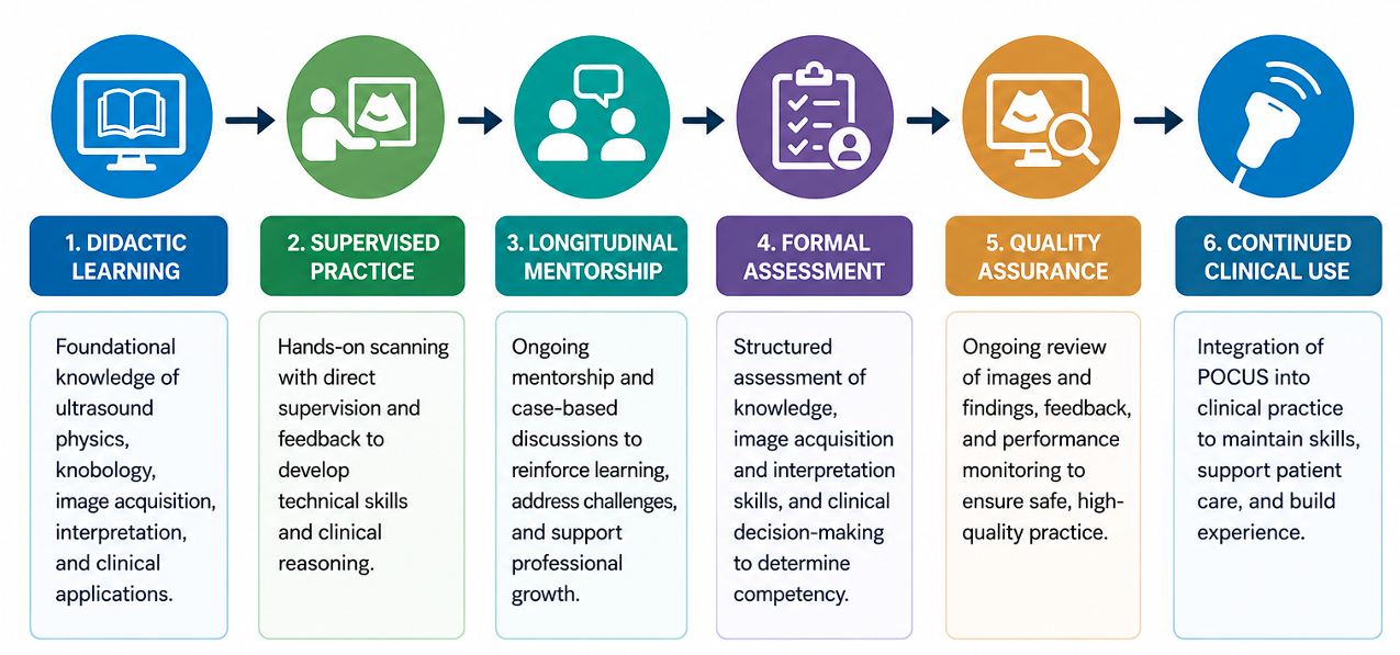 Figure 8. PoCUS training workflow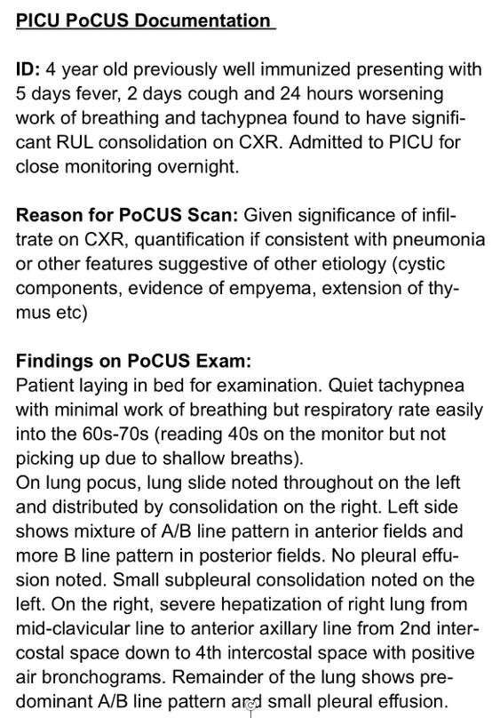
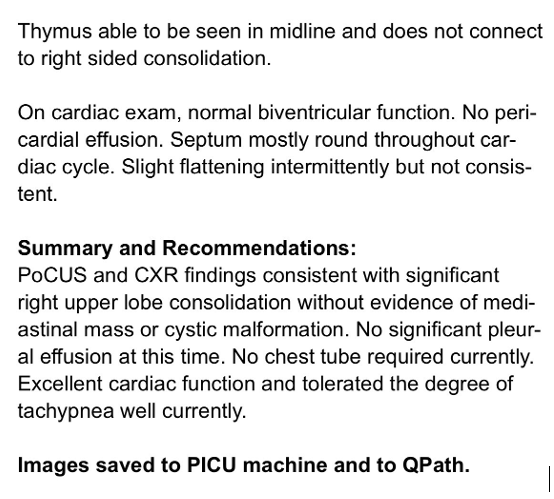 Figure 7. Example of a thorough chart documentation after PoCUS exam
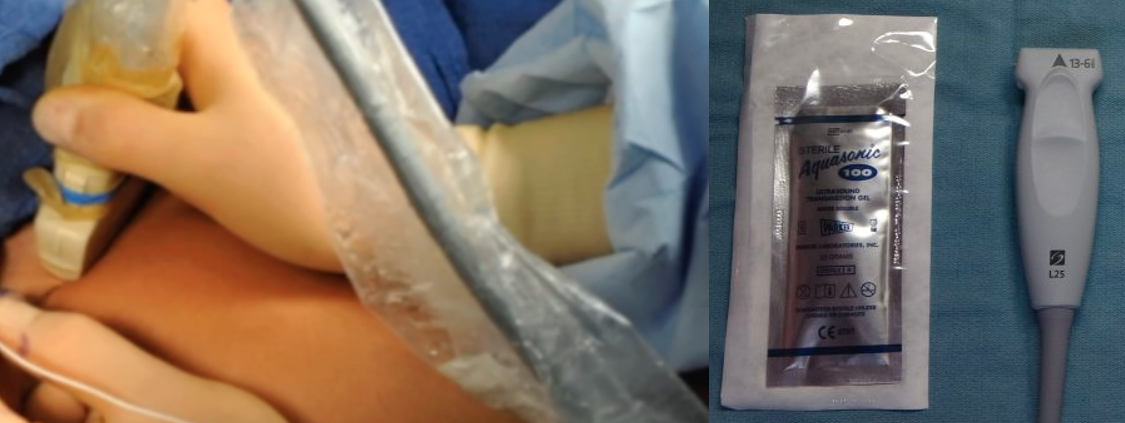 Figure 5. (a)Probe cover and (b)sterile gel
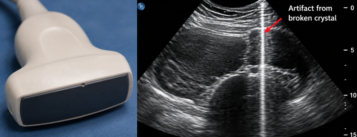 Figure 6. A ) Damaged probe and B) Imaging artifact from broken crystal