Indications
- Dyspnea
- Cough
- Monitoring for volume overload
Equipment
- Ultrasound machine
- High frequency linear probe (6-12 MHz) is most used, although a curvilinear may be better especially in older patients. A phased array probe may also be used.
- Gel
Technique
- Position the patient: It is easiest to completely expose the thorax. LUS can be conducted with the patient in any number of positions; in the parent’s arms (helpful for posterior exam), seated on or lying in a stretcher (lateral decubitus can be used to examine the posterior chest).
- Warm the gel if possible: Younger or sleeping patients may respond better if the gel is warmed. Consider warming the gel between your gloved hands and applying a layer of warmer gel to the chest.
- Scan the patient:
- Set the depth to between 5 – 10 cm, choosing the shallowest depth that gives an appropriate image to maximize resolution.
- Orient the probe marker to the patients’ head and identify the pleural line deep to the ribs in the longitudinal orientation. Ensure the probe is perpendicular to the chest wall to give the clearest image of the pleural line possible.
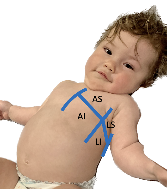
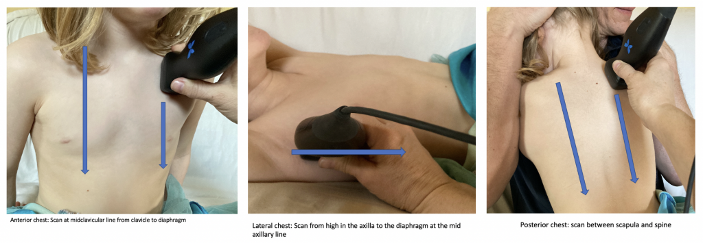
- Scan the patient’s chest systematically: this can be done by dividing the chest into anterior and lateral areas and by investigating each zone superiorly and inferiorly (figure 1). Alternatively, you can choose to investigate each rib space by sliding the probe from cranial to caudal until the diaphragm is reached in each zone as you would in pneumonia, including the posterior zone (figure 2).
- For any abnormalities detected, the area should be investigated fully by sliding the probe along the rib space or changing the probe to a transverse view to scan in-between the ribs.

Figure 1: 8 zone technique for interstitial disease

Figure 2: Alternative technique similar to pneumonia
Tips:
- It may be easiest to position the child seated in a parents’ arms
- For lateral and posterior views lifting the arm or crossing arms in front of the body can allow greater access to the chest superiorly
- Consider warming the gel
- Consider the size of the patient when selecting a probe
- Consider using the “lung preset” if available on your US machine
