
PoCUS findings should be documented clearly, concisely, and in a way that directly answers the clinical question being asked. Documentation should communicate what was examined, what was found, and how those findings influenced clinical decision-making [7]. The goal is not to provide a comprehensive radiology report, but rather a focused assessment that reflects the scope of the examination performed. Appropriate documentation and image archiving are essential components of safe, accountable PoCUS practice.
Use Clear, Binary Language
Whenever possible, PoCUS findings should be documented using simple, binary statements that answer the clinical question.
Examples:
· No pericardial effusion identified.
· Large right pleural effusion present.
· No sonographic evidence of hydronephrosis.
·Qualitatively normal biventricular cardiac function.
This approach reduces ambiguity and aligns with the focused nature of PoCUS examinations.
Link Findings to the Clinical Question
Documentation should clearly connect the examination findings to the reason the scan was performed.
Examples:
· Clinical question: Is there a large pericardial effusion contributing to hypotension?
· Documentation: No pericardial effusion identified.
· Clinical question: Is pulmonary edema contributing to dyspnea?
· Documentation: Bilateral diffuse B-lines identified, consistent with an interstitial syndrome.
Acknowledge Uncertainty and Limitations
Not every examination provides a definitive answer. Poor acoustic windows, patient factors, or limited views may reduce diagnostic confidence. When uncertainty exists, it should be documented clearly.
Examples:
· Study limited by child cooperation.
· Image quality suboptimal.
· Unable to adequately visualize the appendix.
· Indeterminate study. Formal imaging required.
Documenting limitations is a strength, not a weakness. Recognizing uncertainty helps prevent overinterpretation and reduces the risk of false reassurance.
Avoid Overgeneralization
PoCUS is designed to answer focused clinical questions and may not reliably exclude all pathology. Documentation should reflect the scope of the examination performed. A negative PoCUS finding does not necessarily exclude disease.
Avoid statements such as:
“Normal abdomen.”
“No intra-abdominal pathology.”
“Heart normal.”
Instead, document only what was assessed:
No free fluid identified on FAST examination.
No large pericardial effusion identified.
No hydronephrosis visualized.
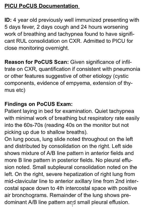
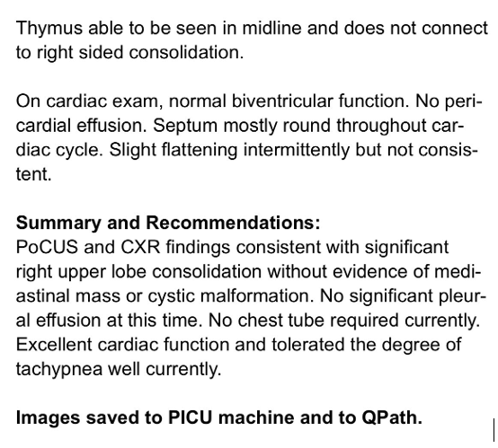 Figure 7. Example of a thorough chart documentation after PoCUS exam
Quality Assurance, Image Archiving, and Accountability
Whenever possible, PoCUS examinations should be archived and available for review. Image archiving supports quality assurance processes, facilitates feedback and education, allows findings to be reviewed in the context of future patient care, and has medico-legal considerations [7]
Saved images should include any relevant labels (e.g left/right, affected/unaffected), image orientation (transverse/longitudinal).
Clinicians remain accountable for the interpretation and documentation of PoCUS findings. Documentation should accurately reflect the examination performed, the findings obtained, and any limitations that may affect interpretation.
For additional guidance on image labeling, image saving and archiving workflows, please revisit the KidSONO Knobology module content and instructional videos
Despite the importance of PoCUS documentation and image archiving, common barriers include perceived time constraints, lack of certification, and concerns about litigation [6]. These perceived barriers should NOT prevent clinicians from chart documentation and archiving representative images, as both are essential components of quality assurance, patient care, and professional accountability.