
LP is performed below the termination of the spinal cord, which in pediatric patients typically ends at the L2–L3 level. The dural sac continues caudally to about S2, making the L3–L4 or L4–L5 interspaces the preferred sites for needle insertion. PoCUS helps confirm the appropriate level and visualize midline structures, interspinous spaces, and depth to the ligamentum flavum–dura complex.
In neonates and infants, incomplete ossification of vertebral elements and abundant cartilage provide excellent acoustic windows, allowing visualization of neural structures that are more difficult to see in older children. These include the posterior CSF (subarachnoid) space, cauda equina, and occasionally the filum terminale.
Sagittal (longitudinal) view
In the sagittal orientation, the vertebral column is viewed longitudinally, allowing appreciation of the alignment and spacing between vertebral elements.
Key anatomical structures include:
· Spinous processes– midline bony projections extending posteriorly from each vertebra.
· Interspinous space– the interval between adjacent spinous processes through which the needle passes.
· Ligamentum flavum– a thick elastic ligament joining adjacent laminae and forming the posterior boundary of the epidural space.
This plane provides an overview of vertebral alignment, and the ligamentous pathway traversed during needle insertion.
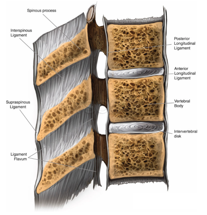 Figure 3. Sagittal spinal ligament and spinal anatomy. Image reproduced with permission from NYSORA (www.nysora.com). All rights reserved.
Transverse View
The transverse view provides a cross-sectional representation of the lumbar anatomy and is used to confirm midline alignment.
Key anatomical structures include:
· Spinous process– midline bony projections extending posteriorly from each vertebra.
· Laminae– paired bony plates extending from the pedicles to meet at the midline, enclosing the vertebral arch.
· Transverse processes– lateral projections serving as muscle attachment sites.
· Ligamentum flavum– positioned deep to the laminae, anterior to the epidural space and dura mater.
· Vertebral foramen– central canal containing dura mater, subarachnoid (CSF) space, conus medullaris, and cauda equina
The transverse view illustrates the symmetry and depth of the posterior elements and helps contextualize the relationship between bone, ligament, and the spinal canal.
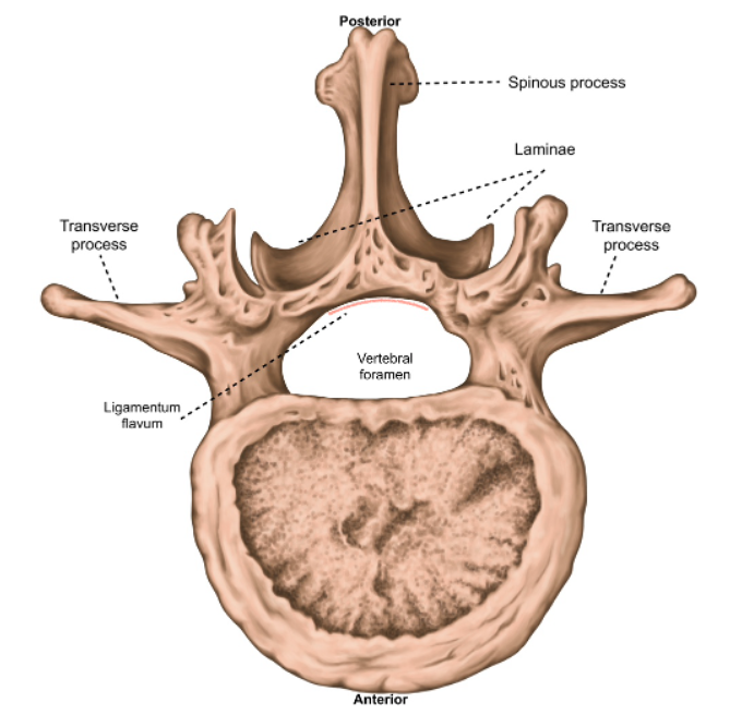 Figure 4. Transverse view of the vertebrae – labeled