What is NOT Normal
Primary Findings:
Ultrasound findings in acute appendicitis typically include [16,17]:
*Most typically a total diameter of >6mm has been used, however it is important to note that there is variability in normal appendix size and some institutions have had success with adjusted criteria to increase specificity and therefore decrease the negative laparotomy rate [18]
** In cases of acute appendicitis, the appendix typically does not compress under probe pressure [19]. However, if perforation has occurred, the appendix may appear compressible, which can complicate the assessment [20]
Figure 9: Inflamed appendix long axis with fluid within the lumen and surrounding hyperechoic fat. Video courtesy of Dave Kirschner, used with permission
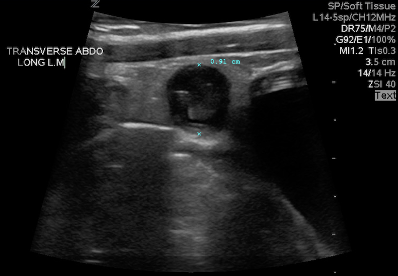
Figure 10: Inflamed appendix in short axis. Image courtesy of Dave Kirschner, used with permission
Figure 11: Easily visualized round, thick-walled structure – Cross section of inflamed appendix. Video courtesy of Dave Kirschner, used with permission.
Secondary/Supportive Findings
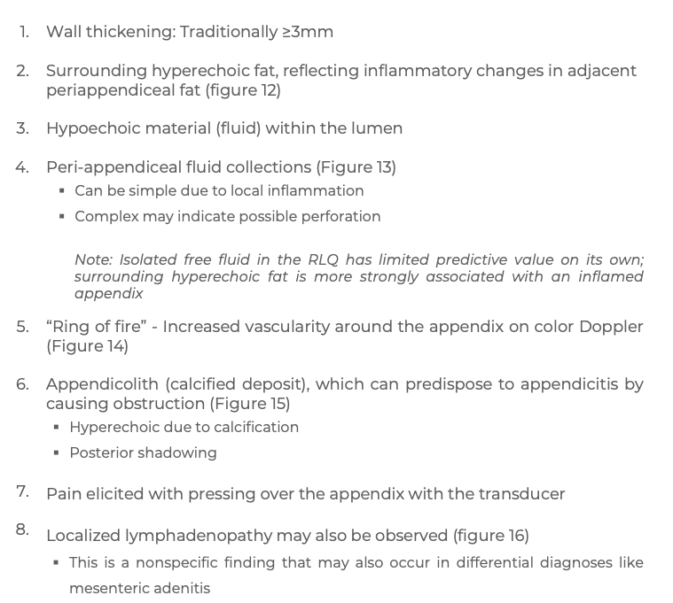

Figure 12: Hyperechoic, reactive fat surrounding the inflamed appendix. Borbély Márton, CC BY-SA 4.0 via Wikimedia Commons [22]
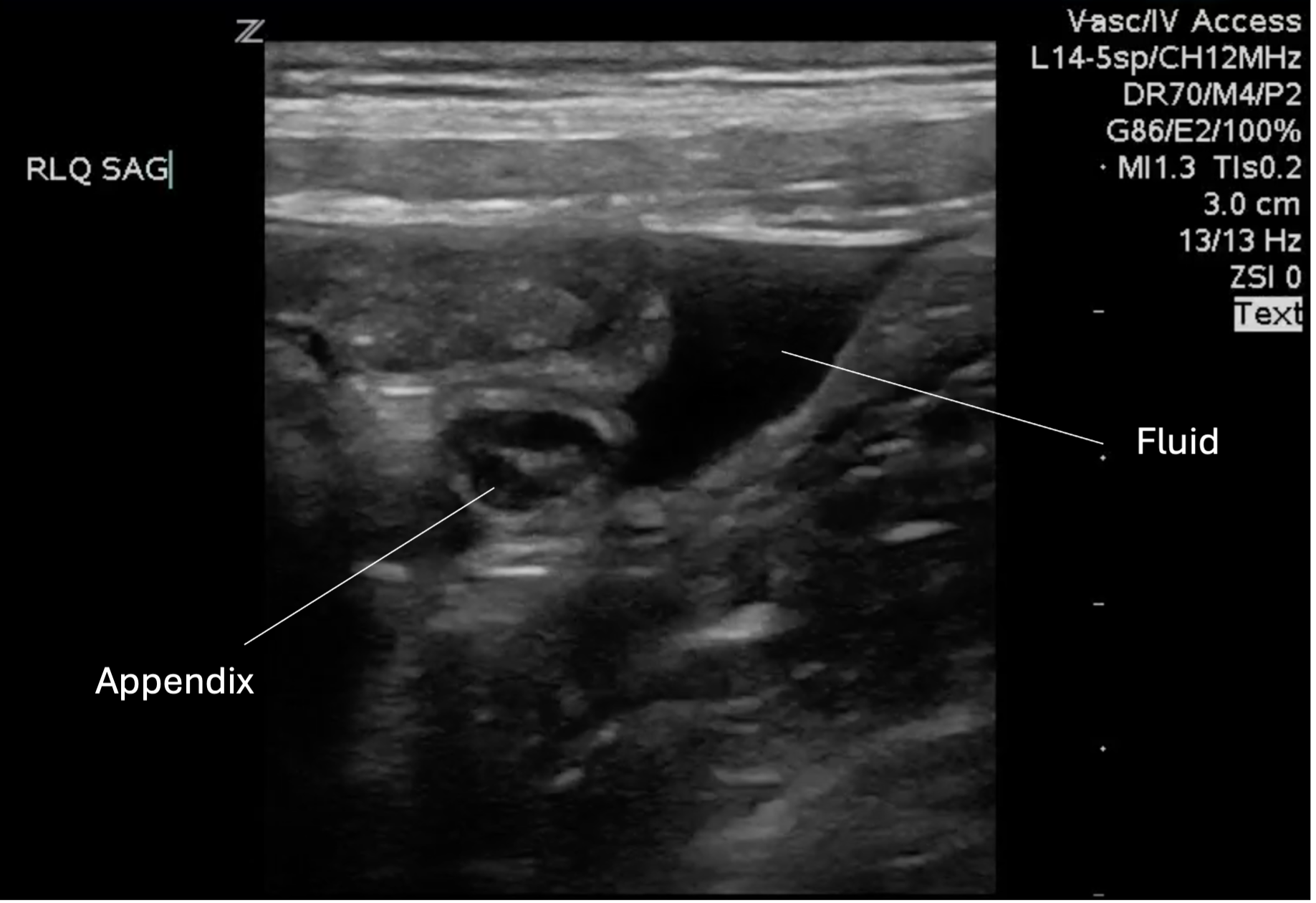
Figure 13: Peri-appendiceal simple fluid collection
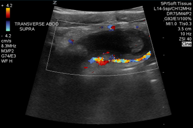
Figure 14: Inflamed appendix with color doppler displaying the “ring of fire” appearance. Image courtesy of Dave Kirschner, used with permission.
Figure 15: Inflamed appendix with appendicolith. Video courtesy of Dave Kirschner, used with permission.
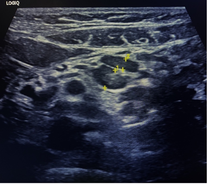
Figure 16: Localized lymphadenopathy at the RLQ