Ultrasound anatomy: What is normal?
First it is important to identify the cecum in the right lower quadrant as the beginning of the large bowel. To identify the cecum first place the probe in the RLQ and identify the psoas muscle and iliac vessels in cross section [Figure 4]. Lateral to the psoas muscle the cecum can be identified as a gas or stool filled structure [figure 5].
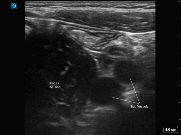 Figure 4: Transverse image of the RLQ, showing the iliac vessels in cross section and psoas muscle
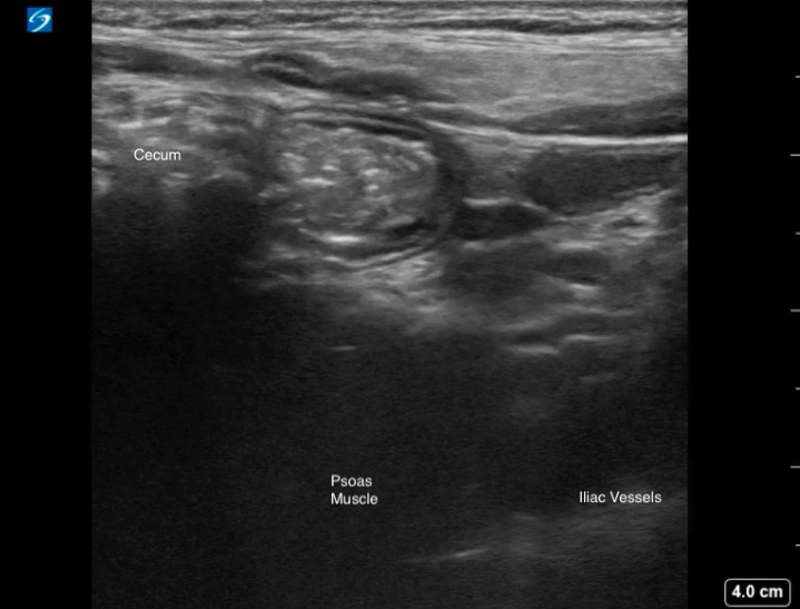
Figure 5: Transverse image of the RLQ demonstrating a gas-filled cecum with posterior shadowing, positioned lateral to the psoas muscle.
Video 1: Transverse scan through the RLQ, displaying the gas filled cecum laterally and normal appearance of the terminal ileum with a smaller luminal caliber.
On ultrasound, distinguishing between small and large bowel can sometimes be challenging. Typically, small bowel appears as having a smaller luminal caliber, contains fluid contents within, and demonstrates active peristalsis. Whereas large bowel has a larger calibre, is typically filled with gas or air and stool, has haustral folds and lacks peristalsis (Video 2).
Video 2: Normal large bowel, displaying haustral folds and lack of peristalsis