What is normal?
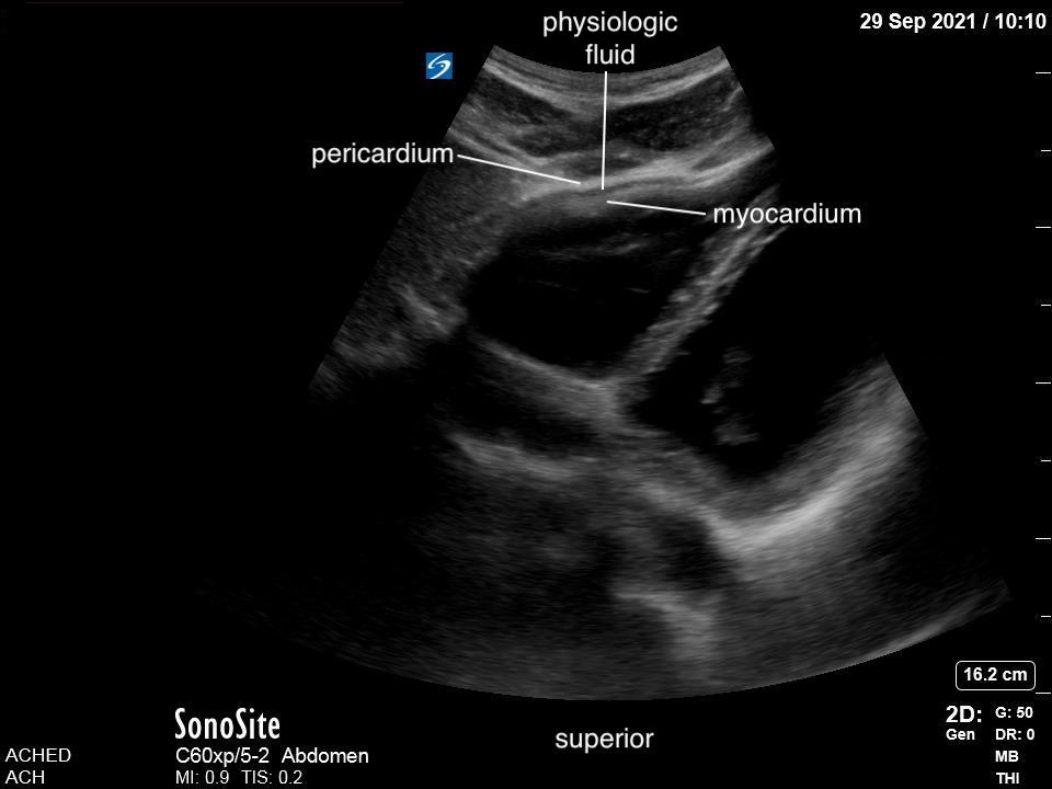
Our area of interest is the space between the pericardium and the myocardium of the left ventricle, extending to the apex of the heart. It is normal for some patients to have visible trace pericardial fluid, which will act as a lubricant for cardiac motion. This, however, will be minimal.

FIGURE 8: Subxiphoid. Physiologic free fluid.

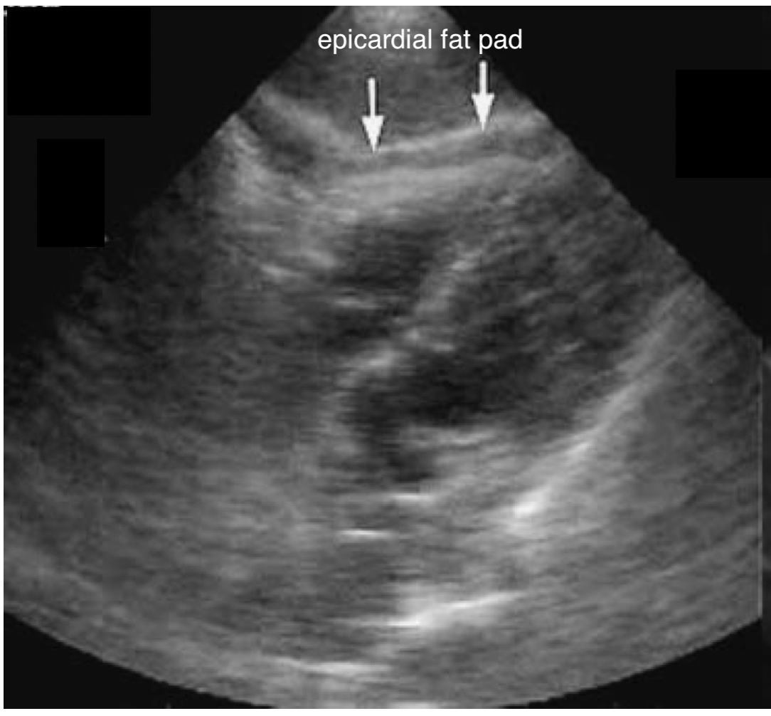
FIGURE 9: Subxiphoid. Pericardial fat pad.
It is also normal for some patients to have a pericardial fat pad. This fat pad sits between the pericardium and the myocardium and has a similar, black, appearance. Unlike fluid, however, the fat pad will be more obvious anteriorly, and disappear as you move posteriorly. Unlike fluid which will move posteriorly to the most dependent area. Also, the fat pads will have very little variation with contraction, while pericardial effusions will show significant variation with contraction.
