What is NOT normal?
There are three characteristic features of a pleural effusion on ultrasound:
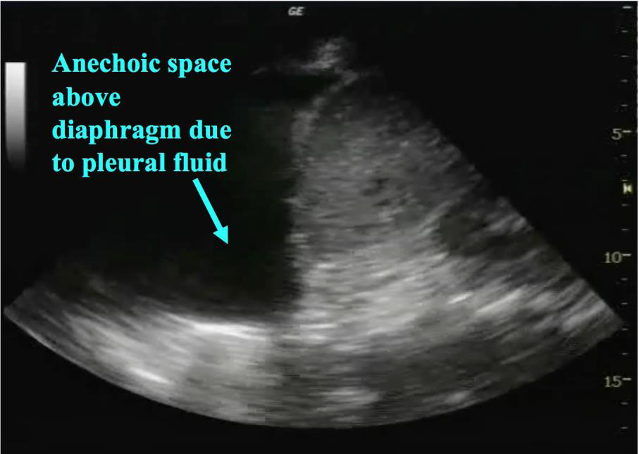
- Anechoic area above the diaphragm
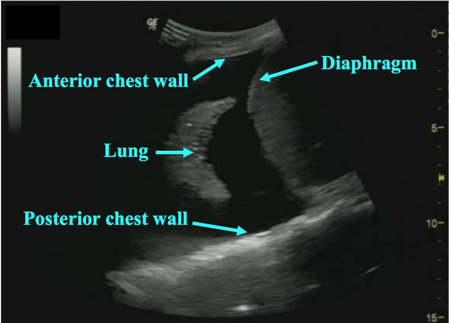
- Anatomic boundaries: diaphragm, chest wall, and lung
- Dynamic movement
Anechoic space:
As fluid collects in the pleural space it can be directly visualized by ultrasound as an anechoic area. The presence of a pleural effusion results in the loss of normal lung artifact: mirror image of the liver if viewed via the abdomen or of A and B lines if viewed via the thorax. The appearance of the fluid itself can vary depending on the type of effusion.
 Figure 7: Anechoic space
Anatomic Boundaries:
Given the fluid in a pleural effusion is not collecting in the lung itself but in the potential space between the chest wall and lung it has distinct anatomic boundaries including (figure 8):
- Chest wall
- Lung
- Diaphragm
 Figure 8: Anatomic boundaries
It is important to note these anatomic landmarks to ensure the location of the fluid, especially prior to performing procedures.
Fluid is an efficient transmitter of ultrasound waves and thus a pleural effusion allows visualization of structures not normally seen. The diaphragm can be visualized with ease and in large effusions can be seen through its entire course. Additionally, the vertebral bodies previously obscured by aerated lung will be visible from the posterolateral approach. This is known as the “spine sign” (video 3).
Video 3: Spine Sign
Dynamic Movement:
Fluid collected in the pleural space is free-flowing in the absence of loculations and adhesions. It subject to the forces of a moving diaphragm, heart and lung. When viewing an effusion via ultrasound it one can note how the fluid conforms to the shape of moving structures (video 4).
Of note, since fluid separates the visceral and parietal pleura normal lung sliding and pleural motion will be eliminated at the level of the effusion as well.
Video 4: Dynamic Movement
Transverse Pleural Effusion Assessment
Transverse evaluation of a pleural effusion offers clinicians added insight into pleural effusions by outlining both the size and characteristics of the fluid pocket, which is useful in guiding decisions around drainage planning.
Video 5: Transverse assessment of a pleural effusion from the axilla to the diaphragm. Rib shadows are visible as the probe passes over each rib during the downward slide. The transverse assessment ends when the diaphragm or spleen/liver comes into view
