Skin and Soft Tissue Ultrasound: A Stepwise Approach
Indications
- Evaluate soft tissue skin infection
- Differentiate cellulitis from abscess
- Evaluate for features of necrotizing fasciitis
- Identify foreign bodies
Equipment
- Ultrasound machine

- High-frequency linear probe (8-12 MHz)
- Water bath or step off pad – if needed
- Sterile probe sheath –if needed
Patient Positioning
- Place the patient in a position of comfort and expose the area of interest.
- Palpate the area of interest ± mark any areas of maximal tenderness or fluctuance.
- Consider allowing access to the opposite side as a “normal” comparison
Ultrasound Machine Preparation
- Ensure the ultrasound screen is positioned in order to be clearly seen.
- Select the high-frequency linear ultrasound probe.
- Consider whether use of a sterile sheath is clinically indicated.
- Choose a vascular or soft-tissue pre-set on the ultrasound machine.
- Select depth of field to between 4-6 cm.
- Focus gain to clearly visualize the soft tissue structures of interest.
Scanning Principles
- Place the linear probe gently over the area in question.
- Adjust the depth and optimize the gain to ensure that the fascial plane is visualized.
- Slowly and systematically scan across the area of interest.
- Scan in both the transverse and longitudinal planes in order to not miss any findings.
- Use color doppler to help identify and differentiate vascular structures
Clinical Pearl: The Dead Zone
The “dead zone” prevents visualization of superficial structures in the near field. This is caused by the ultrasound machine’s inability to send and receive sound waves simultaneously. The depth of the dead zone decreases as frequency increases, and with higher quality send/receive function of the machine and probe. As technology improves the dead zone becomes less clinically significant.
| Tips and Tricks: “What if my area of interest is very superficial?” | |
|
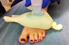
A water bath or standoff pad (such as an IV fluid bag or glove filled with water) can be used to alleviate patient discomfort and improve image quality of very superficial objects. Ensure that the probe is perpendicular to the skin at all times as an oblique angle can cause false hypoechogenicity.
Note: A recent study showed that although most (89%) new ultrasound probes have a “dead zone” of 0 mm, older probes have a “dead zone” of up to 3 mm [13]. |
|
| Water Bath | Standoff Pad |
 |
 |
Video 1: Video of normal finger in a Waterbath
From EDSonoshare Library
