The echogenic capsule makes the bean-shaped organ easily identifiable. The cortex is either isoechoic or hypoechoic (more commonly) compared with the normal liver or normal spleen. However, in neonates and young infants, the renal cortex may appear more echogenic than the liver, which is a normal age-related finding. The renal cortical tissue extends into the medulla, separating the pyramids in the form of columns called columns of Bertin. The medullary pyramids are hypoechoic or anechoic compared to the cortex. On most occasions, the pyramidal shape is not well visualized but is often better seen in small children. Renal sinus fat is echogenic and occupies a major part of the inner kidney. The collecting system is usually not visualized unless distended and is embedded in the surrounding echogenic sinus fat. The renal pelvis area is hypoechoic but not “black” unless there is hydronephrosis. Similarly, ureters are not typically seen unless distended as in hydroureter [13].
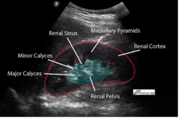
Figure 9: Normal right kidney in longitudinal view with labeled renal anatomy. Image use with permission POCUS 101 [10]
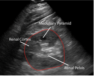
Figure 10: Normal left kidney in longitudinal view with labeled renal anatomy Image use with permission POCUS 101 [10]