The shape and relationships of the bladder on ultrasound examination will depend on its degree of filling. The sagittal view is triangular (figure 10), and the transverse view is rectangular (figure 11). That said, the shape varies depending on bladder fullness. When distended, the bladder walls appear thinner compared to a thicker appearance when less distended (figure 12).
No single cutoff defines a normal bladder capacity or normal PVR across all pediatric patients due to variability with age and gender. However, as a general rule of thumb, a PVR less than 10% of the estimated bladder capacity is considered normal [12-14].
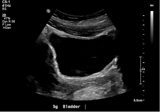 Figure 10: Normal bladder in the sagittal plane
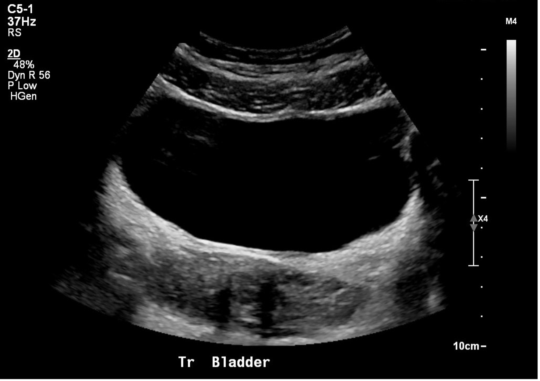
Figure 11: Normal bladder in the transverse plane
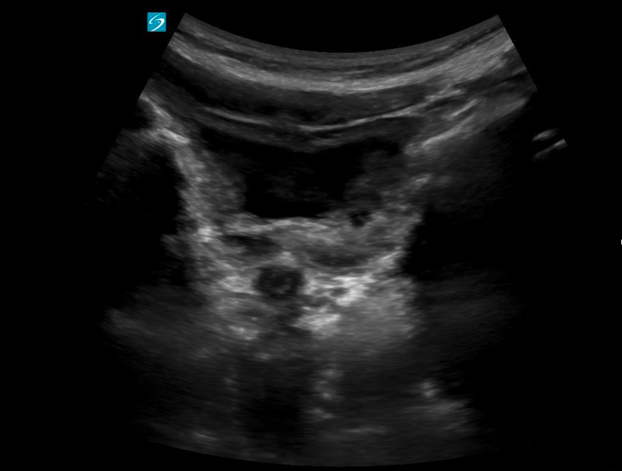 Figure 12: Transverse view of a near empty bladder with thicker walls
Catheterized Children
In catheterized children with a Foley in place, the bladder may appear partially or completely decompressed depending on recent drainage. The Foley catheter is typically visualized as a linear echogenic structure within the bladder lumen, often with posterior shadowing. The inflated balloon at the catheter tip appears as a well-defined, round anechoic or hypoechoic structure with an echogenic exterior rim, centrally located within the bladder (Figure 13).
Note: The presence of internal debris within the bladder lumen (seen as echogenic material or echoes within the fluid) may be considered a normal finding in patients with long-term catheter use.
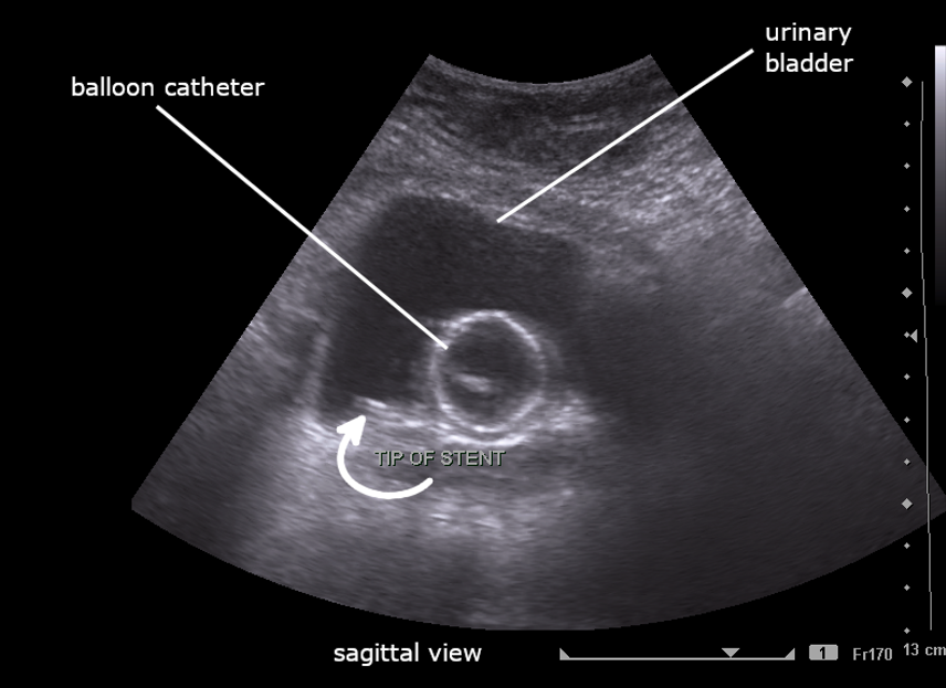
Figure 13: Sagittal image of partially filled bladder with Foley catheter. Image By Cerevisae – CC BY-SA 4.0 license.
Figure 14: Transverse view of a fully decompressed bladder with a normal foley balloon. Video courtesy of Dr. Dan Kim, used with permission.