Orientation
When scanning the bladder, like with any ultrasound, the transverse plane provides a cross-sectional view, with patient right on the screen’s left, patient left on the screen’s right, and anterior in the near field and posterior in the far field. In the sagittal plane, cranial is on the screen’s left, caudal is on the screen’s right, with anterior in the near field and posterior in the far field.
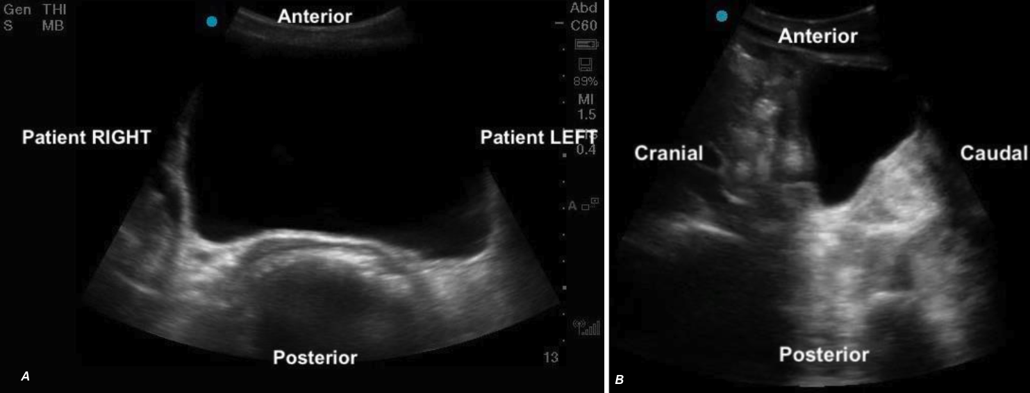 Figure 5ab: Transverse and sagittal bladder views with orientation labels
Appearance:
The bladder will appear as an anechoic cavity surrounded by echogenic walls.
Position
The bladder sits in the pelvis just posterior to the pubic symphysis. Its size and position relevant to other structures varies according to bladder fullness.
> Full Bladder:
– Sits anterior to the uterus in females (figure 7a, 9a)
– Sits cranial and anterior to the prostate in males (Figure 7b,9b)
– Anterior to the rectum
> Partially filled:
– Sits lower in the pelvis, appearing caudal to the uterus (figure 8)
– Remains anterior to the prostate in males but is positioned lower in the pelvis
– Anterior to the rectum (figure 8)
It’s important to be aware that in younger pediatric patients, when the bladder is near empty, surrounding structures such as the uterus or prostate may also be difficult to identify due to their size or degree of development.
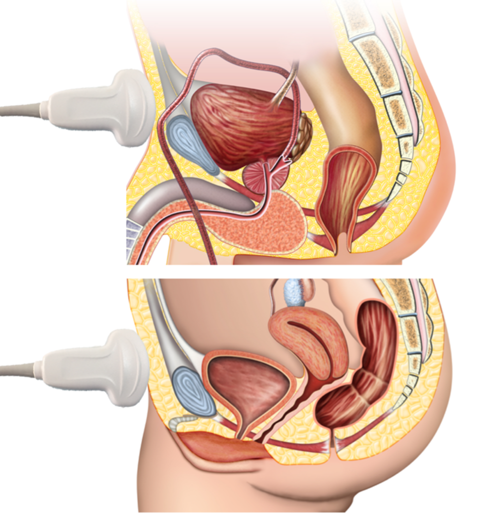
Figure 6: Anatomic diagram of pelvic structures illustrating key landmarks for bladder ultrasound scanning, including the bladder, uterus/prostate & rectum
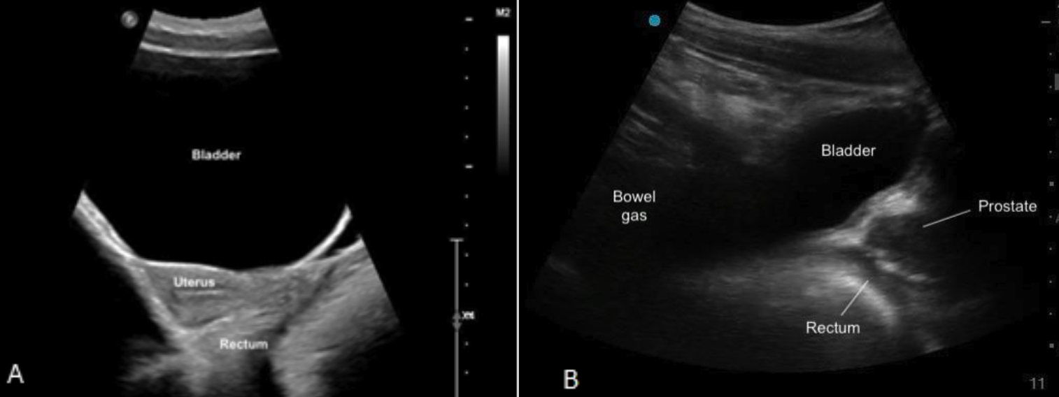
Figure 7ab: Sagittal bladder sonoanatomy in female (a) and male (b) children.
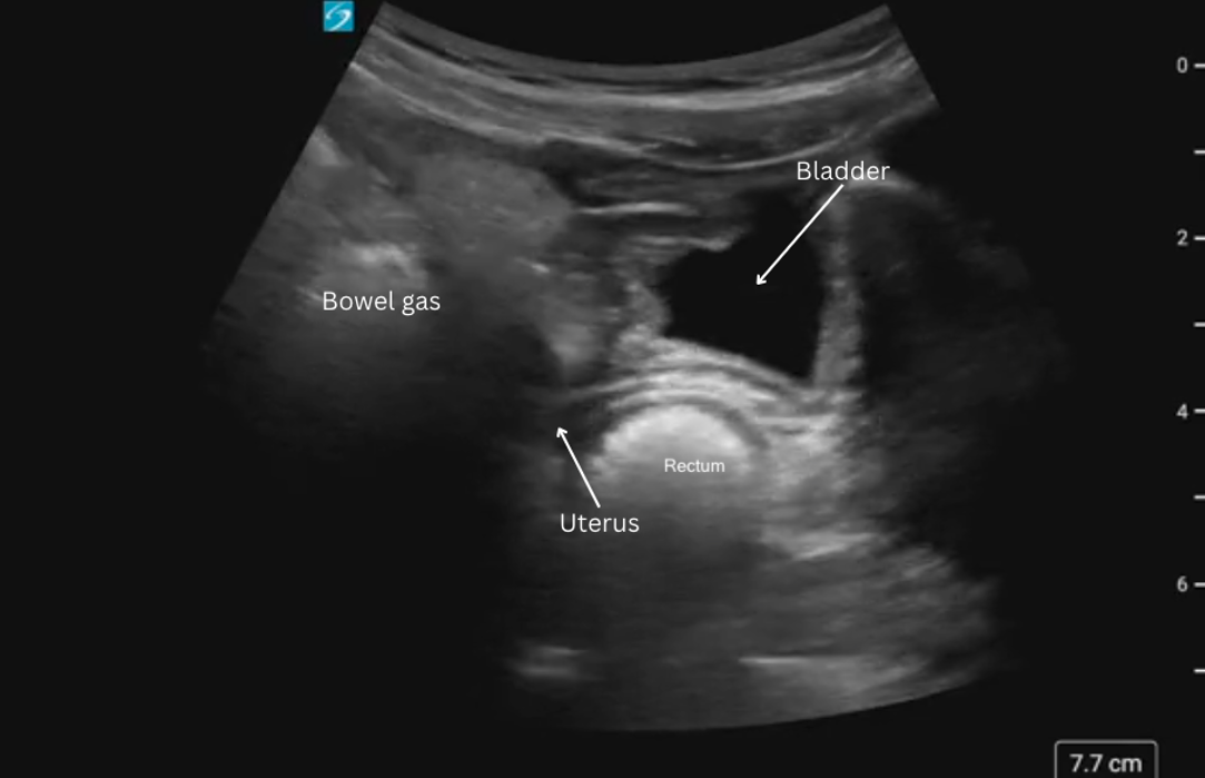
Figure 8: Sagittal view of bladder sonoanatomy in a female patient with a near empty bladder.
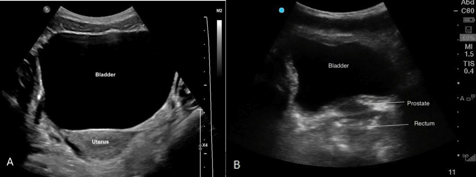
Figure 9ab: Transverse bladder scan sonoanatomy in female (a) and male (b) chidlren.