The RV is typically assessed by dividing it into three key regions: the inlet, the body/apex, and the outlet (infundibulum or conus) (figure 1,2). The inlet includes the tricuspid valve (TV) and the portion of the RV just beyond it. The body/apex represents the most distal and trabeculated portion of the RV. The outlet leads to the pulmonary valve and main pulmonary artery. Because the RV is wrapped around the LV and lies just beneath the sternum, different PoCUS views emphasize different RV regions. Recognizing which regions are visualized in each view is essential to accurately assessing the RV.
The RV free wall refers to the outer wall of the RV that is not shared with the LV, lying opposite the interventricular septum (IVS). The RV free wall is typically described in three segments: basal, mid, and apical (figure 3). These segments are based on proximity to the tricuspid valve and apex. On ultrasound, the RV free wall can be seen in various views (especially the apical 4-chamber and subcostal 4 chamber).
The RV is characterized by prominent trabeculations and contains the moderator band, a muscular ridge crossing the RV cavity that can appear as a distinct linear structure on imaging (figure 3).
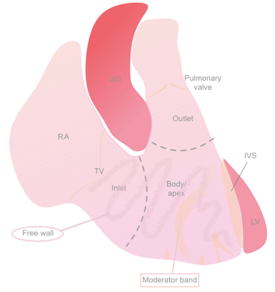 Figure 1: Anatomical diagram of the RV
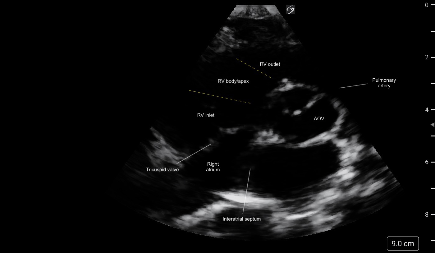 Figure 2: PSAX (aortic level) view illustrating the different regions of the RV and its crescent shape.
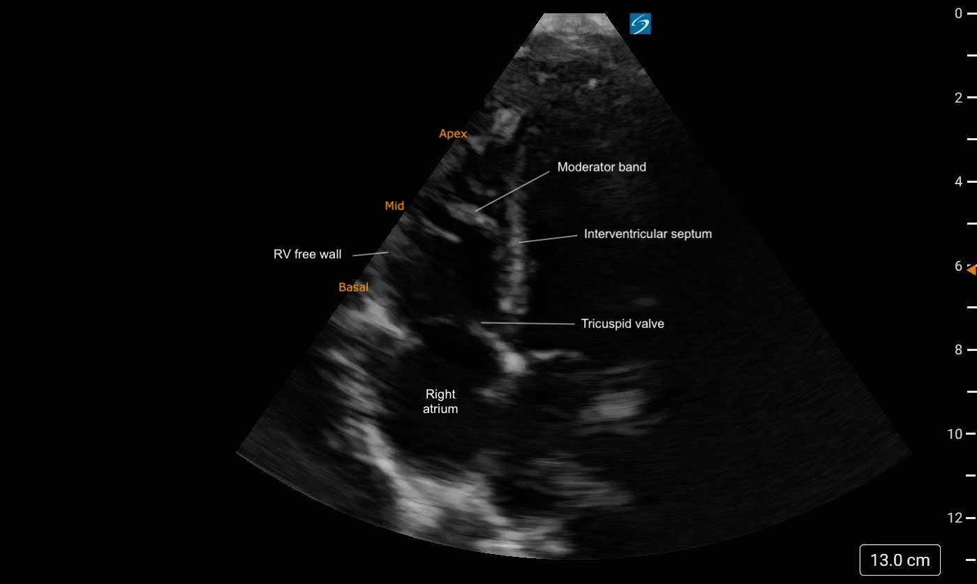 Figure 3: A4C Right heart focused view labeled. Note the trabeculations present at the apex of the RV