Knee Anatomy Review
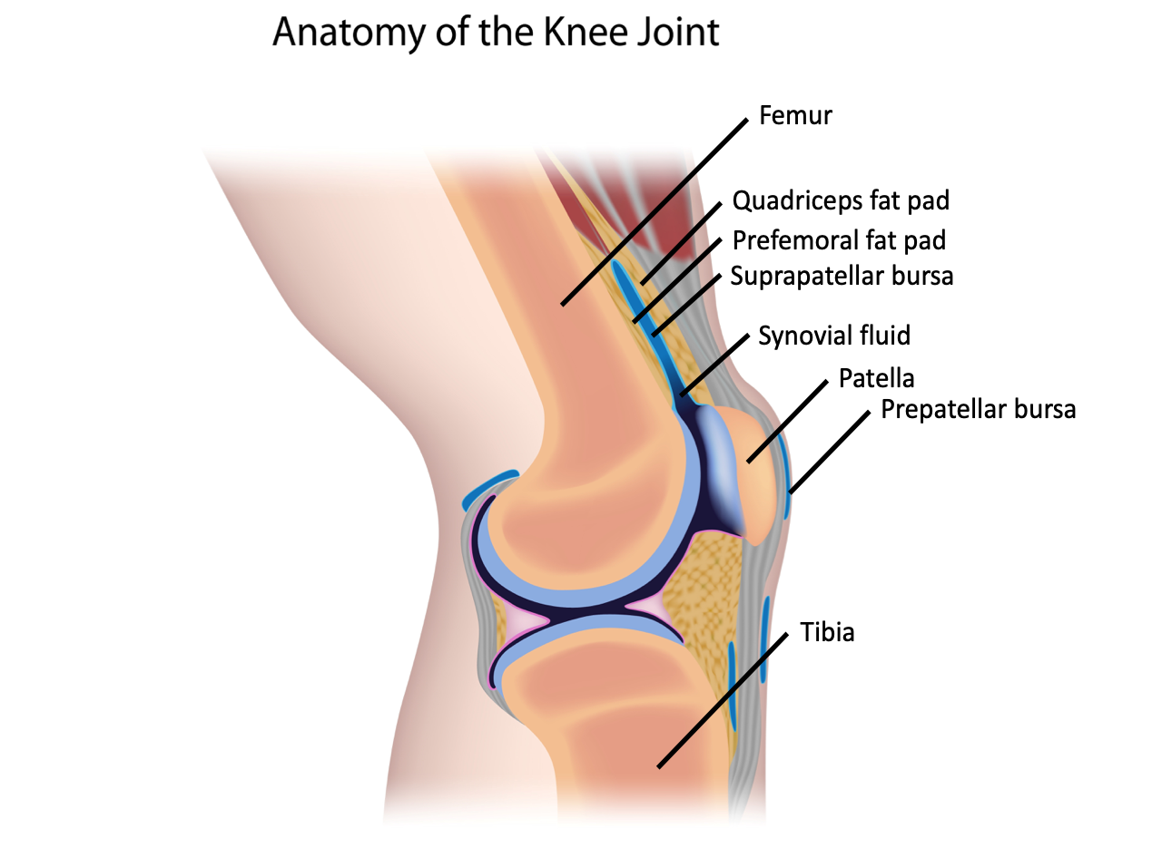
The knee is made up of the articulating surfaces of the femur and the tibia, with the patella lying anteriorly. The quadriceps tendon inserts on the patella, and the patellar tendon extends distally to the tibial tuberosity. The fluid within the synovial capsule is continuous with the suprapatellar bursa. The fat pads found within the knee joint include the quadriceps fat pad and the pre-femoral fat pad (Figure 10).
Figure 10: Anatomy of the knee joint.
Technique
- Position the patient supine.
- Use a towel roll to place the affected knee in 20-30 degrees of flexion [1] (Figure 11).
Figure 11: Probe position for knee ultrasound
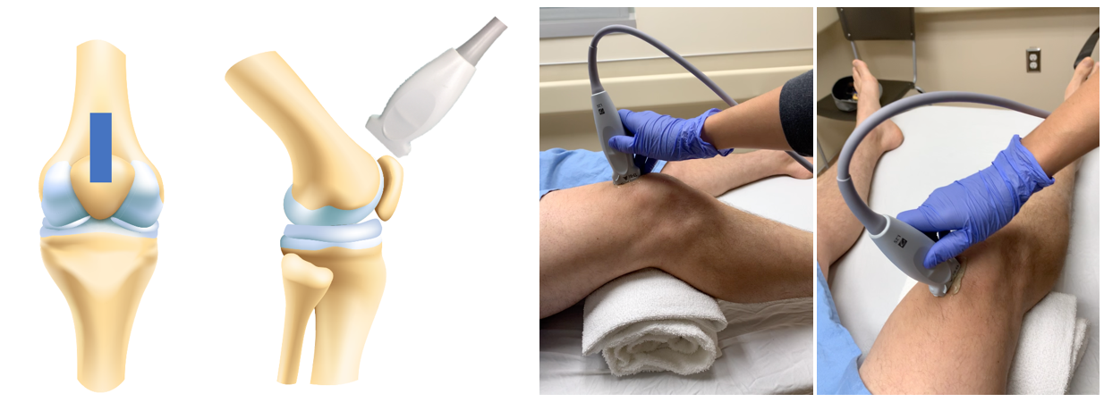
- Place the linear array probe longitudinally in the sagittal plane on the patella [10].
- Identify the patella and scan proximally to assess for effusion within the suprapatellar recess [10].
- Assess for presence of joint effusion.
- Repeat on the contralateral joint.
What am I looking at?
Figure 12: Labelled normal knee ultrasound.
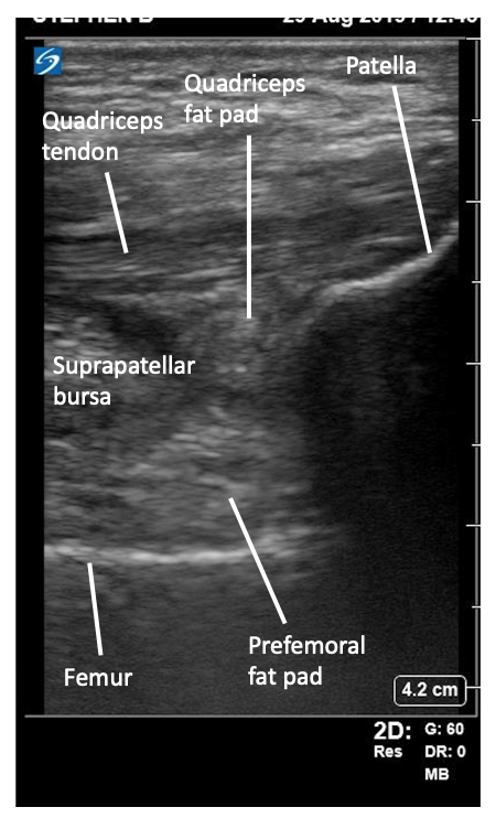
Patella
- Hyperechoic line in caudal field of view.
Femur
- Hyperechoic line in cephalad field of view.
Quadriceps tendon
- Hyperechoic fibrillar structure in near field.
Fat Pads
- Pre-femoral fat pad – hyperechoic soft tissue collection just anterior to femur.
Quadriceps fat pad – hyperechoic soft tissue collection just inferior to quadriceps tendon.
Suprapatellar bursa
- Hypoechoic potential space.
Prepatellar bursa
- Hypoechoic area superficial to patella.
What is normal?
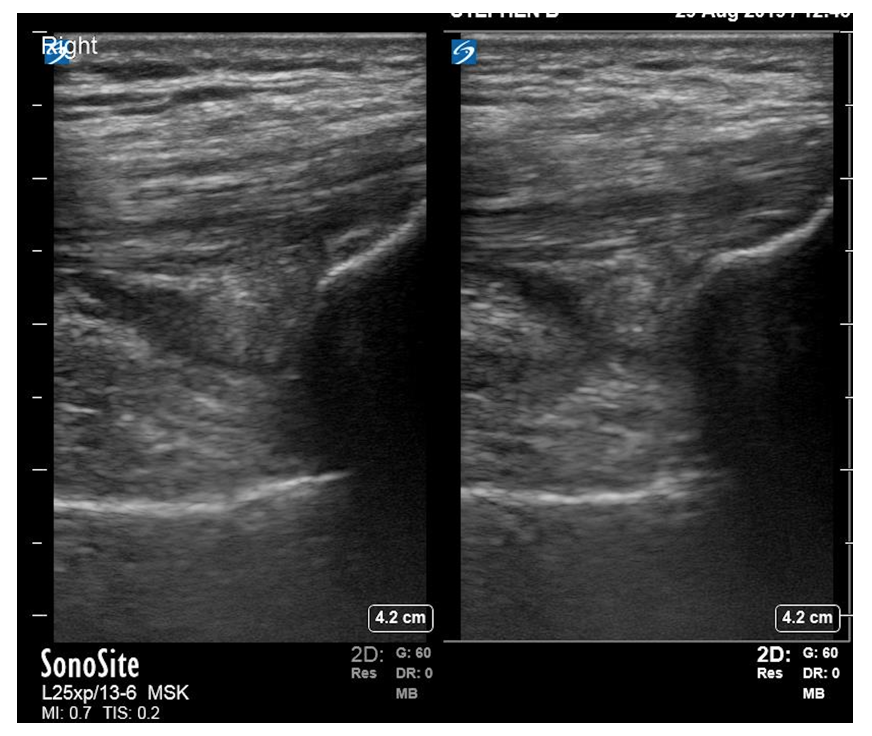
Visualization of the knee joint in this view will show you the patella caudally and the femur extended cranially (Video 2). The quadriceps tendon will be a fibrous structure running in the near field (Figure 13). The suprapatellar bursa should be less than 2mm thick [1].
Video 2: Normal knee ultrasound.
Figure 13: Normal right and left knee ultrasound.
What is NOT normal?
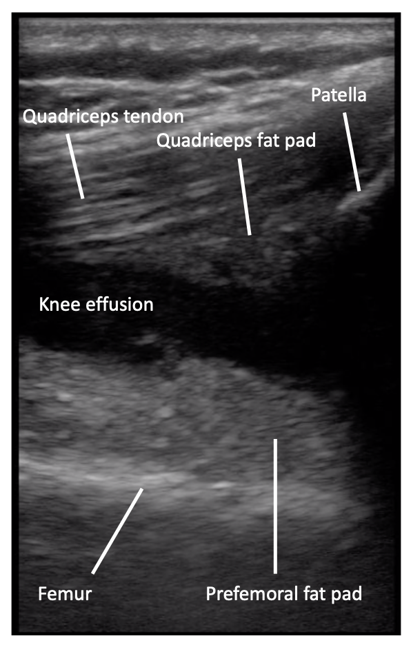
Fluid will collect in the suprapatellar bursa, found in between the prefemoral and quadriceps fat pads (Video 3). The ultrasound is positive for a knee effusion by a collection of hypoechoic fluid which is >2mm thick [1] (Figure 14). If you are not seeing any fluid collection, flexion of the knee can increase the fluid in the suprapatellar recess [13]. In addition, fluid can collect on both the medial and lateral side of the suprapatellar region, therefore scanning these areas can help identify fluid collections. To do this, slide the probe laterally and medially within the sagittal plane to identify hypoechoic fluid collections. This can help with performing ultrasound guided arthroscopy of the knee, although that is outside the scope of this module.
Video 3: Knee joint effusion. Note the fluid collection between the quadriceps fat pad and quadriceps tendon superiorly and the prefemoral fat pad inferiorly.
Figure 14: Positive knee effusion.