Stepwise Technique
Position: Supine or head elevated at 30 degrees, on the bed or in a caregiver’s lap
1. Gather your equipment and select the ocular/ophthalmic preset
2. Have the child close their eye and apply the Tegaderm adhesive (if available).
· If no Tegaderm adhesive is available, it is essential the child keeps their eye closed throughout the exam and to use sterile gel
· Ensure to press firmly at the inner canthus to avoid having air bubble under the Tegaderm
3. Apply a copious amount of sterile gel over the affected eye so the probe floats on the surface, minimizing pressure on the globe.
4. Place the probe in the transverse position over the affected eye (probe marker pointing towards the patients right) (figure 2).
5. Fan the probe superiorly and inferiorly until the anatomy of the eye is clearly visualized
6. Fully scan through the eye from superior to inferior, assessing the anatomy throughout. Document findings
7. Assess extraocular movements – ask the child to move their eyes left and right
8. Rotate the probe 90 degrees clockwise (probe marker facing cranially) to assess in the sagittal plane (figure 3).
9. Fan the probe medially and laterally until the anatomy of the eye is clearly visualized
10. Fully scan through the eye from medial to lateral, assessing the anatomy throughout. Document findings
11. If uncertain of findings, assess the non-affected eye to compare
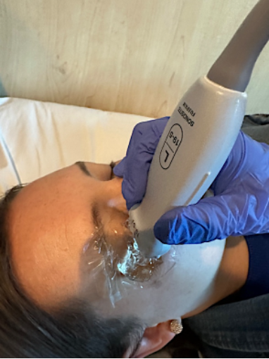 Figure 2. Transverse probe position during ocular PoCUS with Tegaderm and copious gel
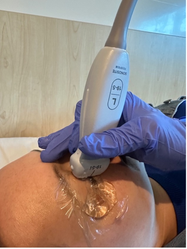 Figure 3. Sagittal probe position during ocular PoCUS with Tegaderm and copious gel