Bladder Scan Technique:
1. The patient will lie supine.
2. Identify the symphysis pubis (figure 1) and place the probe just superior on the lower abdomen with the probe indicator facing patient’s right
3. Identify the bladder

4. Scan thoroughly in the transverse plane by fanning the probe cranial to caudal
5. Rotate the probe 90 degrees clockwise to obtain a sagittal probe position (probe marker pointed cranially).
6. Scan thoroughly in the sagittal plane by fanning through the bladder from hip to hip.
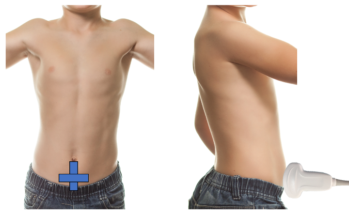
Figure 1. Bladder volume probe positioning
Bladder Volume Technique:
Bladder volume can be determined non-invasively by measuring the bladder in its maximal width, depth, and length. Bladder volume can be calculated both pre- and post-void.
1. Scan the bladder in two planes to obtain three unique measurements (Figure 2)
> Width: In the transverse plane, measure the diameter between the lateral walls to obtain the width.
> Depth (height): is the anteroposterior diameter, which can be obtained in the transverse or sagittal plane. Choose just one plane for this measurement.
> Length: is the craniocaudal diameter, which is obtained by measuring from the superior to inferior wall of the bladder in the sagittal plane.
2. Calculate Volume
> Most current machines contain automated calculators for volume measurement.
– To access the bladder volume calculation package, select the Calculations/Calc button on your ultrasound system. Then search for or navigate to the Bladder Volume option within the calculation menu. Once selected, choose the L, W, or H measurement – as appropriate for the imaging plane you are in.
> As an alternative, the simple formula (LX W X D X 0.7) can be used to estimate bladder volume.
– However, because of the inherent variability of bladder shape and the variation in this shape with differing degrees of filling, bladder volume measurements obtained in this fashion may have an error rate between 15% and 35% [10].
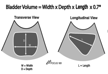
Figure 2: Measurements for bladder volume in transverse and sagittal planes [11]
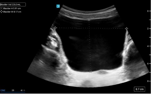 Figure 3.
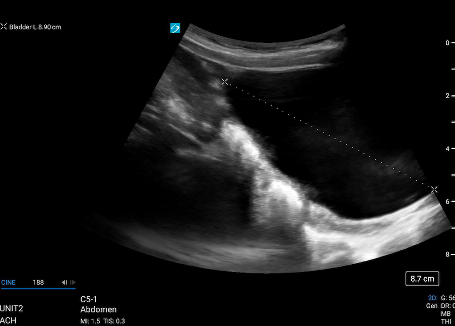 Figure 4.
Figure 3 & 4: Caliper placement for bladder volume in the transverse and sagittal planes.