Pitfalls:
- Failing to fan all the way posterior to include the vertebral bodies—thus missing smaller collections
- Intraperitoneal free fluid: note anatomic landmarks
- Loculated effusions
- Pleural adhesions
- Can’t differentiate the type of fluid (blood vs lymph vs transudate vs exudate)
Characterizing Effusion:
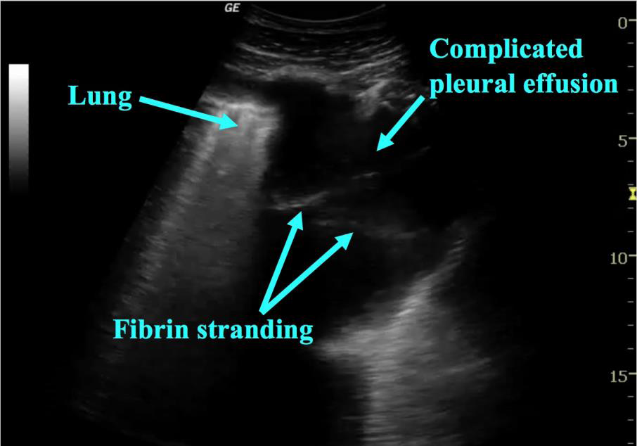
While definitively characterizing effusions is out of the scope of PoCUS it is useful to be aware of how different types of effusion appear on ultrasound. As fluid becomes more complex (clotted blood, pus, adhesions) It becomes more echogenic. Fibrin stranding and loculations appear as echogenic lines of material traversing the fluid (figure 9). [3] If these are seen on PoCUS further imaging or consultation may be required to better characterize the effusion.
 Figure 9: Complicated Effusion
Quantifying Effusion:
While there have been many studies showing complex methods of estimating pleural effusion volumes, we will not review each as this is beyond the scope of PoCUS. Generally speaking, effusions are considered small when the anechoic fluid visualized is limited to the costodiaphragmatic recess and does not extend beyond the dome of the diaphragm. The decision to drain an effusion should be made based on clinical factors in the presence of an effusion, not on the size itself. Additionally, in pediatrics there are no validated, simple ways of estimating volume.
