It is important to visualize an intussusception in both planes to reduce the likelihood of mistaking it for thickened bowel loops, psoas muscle, stool, or other abdominal masses (Figure 11).
Small bowel intussusceptions can often be seen in the abdomen and in most cases are transient and do not require treatment. Small bowel intussusceptions can be differentiated by assessing for size (generally less than 2cm in diameter) and for the presence of peristalsis (absent in ileocolic intussusceptions) (Video 4).
Pathologic lead points may not be easily recognized with POCUS, therefore consider other imaging modalities when patients are outside the typical age range or with suggestive history or physical examination findings.
Ileocolic intussusception may self-reduce, therefore a positive POCUS examination with a subsequent negative radiologist-performed ultrasound is possible.
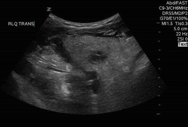
Figure 11: Inflamed, thickened bowel, not to be mistaken for intussusception
Video 4: Small bowel intussusception with the presence of peristalsis