Subxiphoid Four Chamber View
Technique
- In this view, the heart is imaged in a coronal plane but from a different angle than the apical four-chamber view (Fig 21).
- The probe should be held “overhand” with the hand on top of the probe.
- Place the probe in the subxiphoid space with the ultrasound beam pointing up towards the patient’s left scapula.
- In emergency convention the probe marker is pointing towards the patient’s right (figure 22A) and in cardiology convention it is directed towards the left (Figure 22B).
- Depth should be adjusted to ensure visualization of the left ventricle and posterior pericardium
- Gain should be adjusted so the myocardium appears grey and blood black.
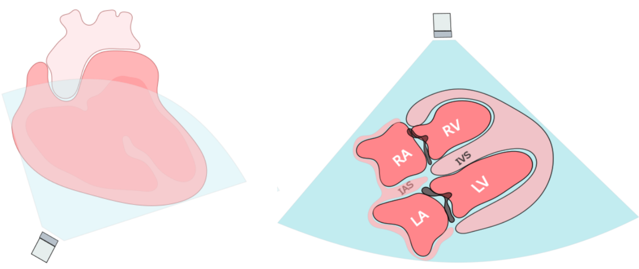
Figure 21: Anatomical coronal view of the heart from the SubXiphoid position
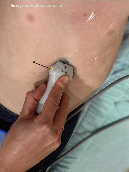
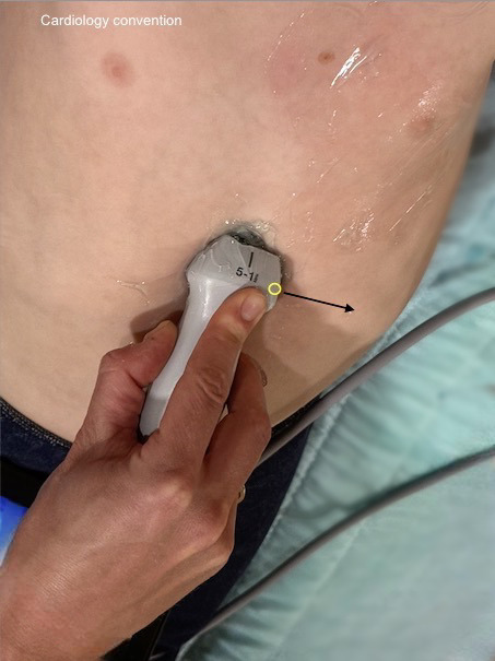
Figure 22. Subxiphoid external landmarking EM vs Cardiology Convention with overhand grip
Note: Emergency vs Cardiology convention
The subxiphoid four chamber view will appear the same on the screen whether you are using emergency or cardiology convention. This is because both the probe markers and screen markers are oriented opposite, resulting in the same net image on screen
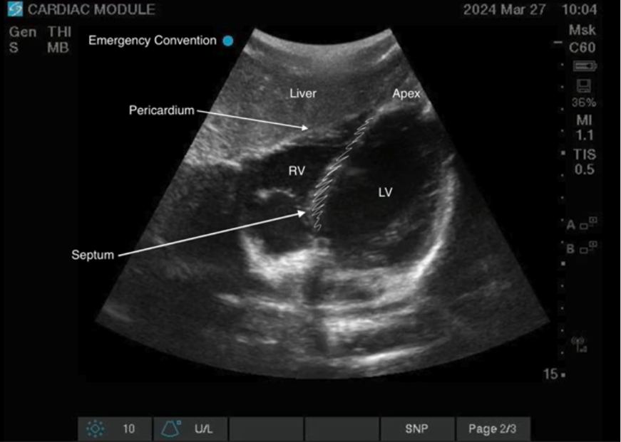
Figure 23: Subxiphoid View EM Convention.
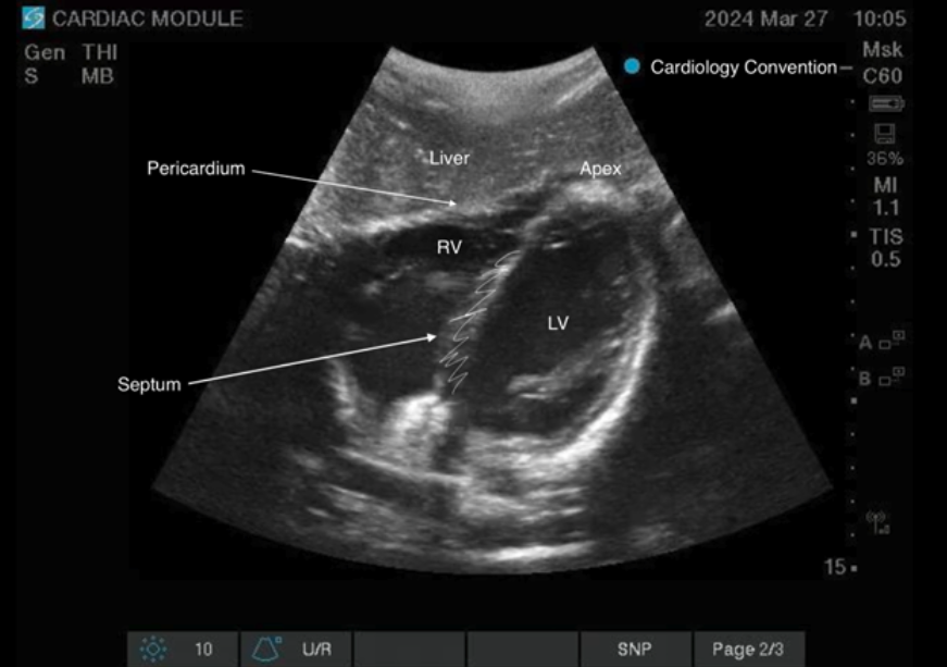
Figure 24: Subxiphoid View Cardiology Convention.
Scanning Tips:
- This window can be challenging to obtain in patients with obesity, abdominal pain or lots of bowel gas as it uses the liver as an acoustic window to view the heart.
- Try using lots of gel and exerting the minimal needed pressure, although sometimes significant pressure is needed.
- If able, having the patient bend their legs can help relax the abdominal wall. Asking the patient to take a deep breathing in can also bring the diaphragm and heart towards the probe.
- If struggling with bowel gas, try moving the probe inferiorly and to the patient’s right to get a window through the liver.
- Fan the probe, flattening it out until the heart comes into view on the screen.
What Am I Looking At?
In this view a coronal view of the heart is obtained. In the near field just below the probe often the acoustic window of the liver can be seen. Just deep to that is the diaphragm upon which the right ventricle lies. Deep to that the left ventricle can be seen. In this view the ventricles are on screen right and the atria are on screen left.
Figure 25: Video of the Subxiphoid 4 chamber view
Clinical Utility
This view is the best view to appreciate pericardial effusions. As it is also a four chamber view it also allows for assessment of the left and right ventricular size and function. Importantly this may be the only view available in emergent situations such as ongoing CPR so as not to interfere with chest compressions.
