Sonoanatomy Review
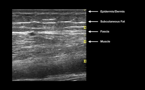
Normal skin and soft tissue have many layers (Figure 1). The most superficial structures are the epidermis and dermis, which appear as if one hyperechoic structure. Deep to this, you will find subcutaneous fat, which is hypoechoic and globular. Blood vessels, nerves and lymph nodes are found within the hypodermis, and are varying in their echogenicity. Deep to these structures, the muscle is found beneath a hyperechoic layer of fascia. Muscle is seen as highly organized hypoechoic and striated fibers.
Figure 1: Anatomy of normal skin
Tendons are visualized as hypoechoic fibrillar organized structures, whereas fat pads are also hypoechoic, but are more homogenous in their appearance (Figure 2). Finally, smooth hyperechoic bone cortex may be seen as the deepest layer.
Figure 2: Anatomy of a normal joint

Tip:
Scan with dual screen functionality
In order to facilitate comparing an affected joint with the contralateral side, it can be useful to use the dual screen function on the ultrasound machine.
- Choose “More Controls” from the main screen, found at the bottom right corner (Figure 3a).
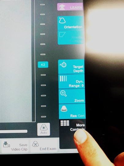 Figure 3a: Choose “more controls”
 - Select “Dual” from the options screen (Figure 3b)
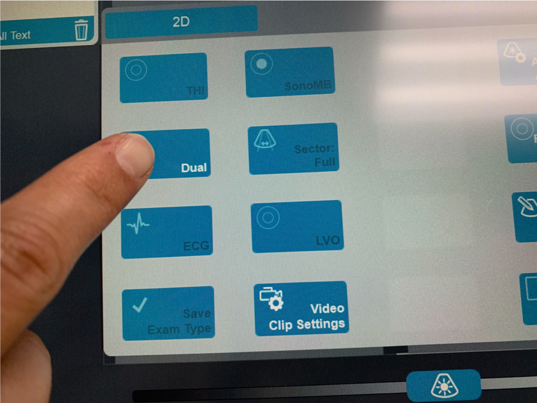 Figure 3b: Choose “dual”
 - Touch the screen on either side to start scanning. When ready to switch, freeze the area of interest on the screen and then touch the opposite screen to begin scanning the opposite side (Figure 3c).
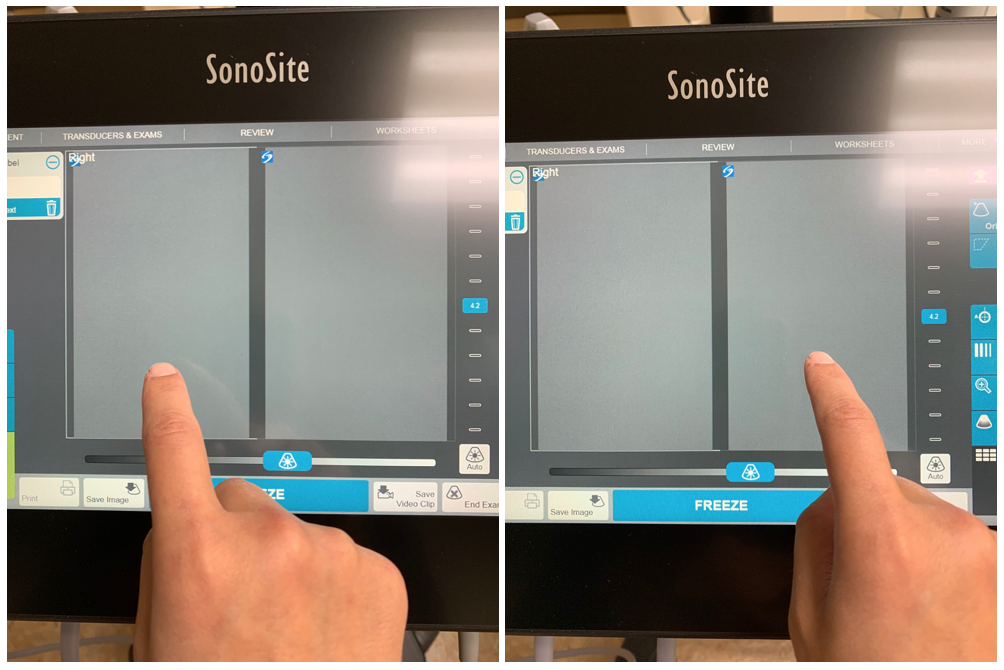 Figure 3c: Toggle between screens by touching the desired side
 - Below is an example of a split screen view (Figure 3d).
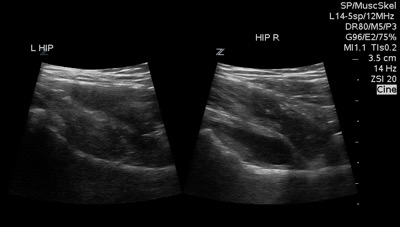 Figure 3d: Dual screen example
