Understanding images
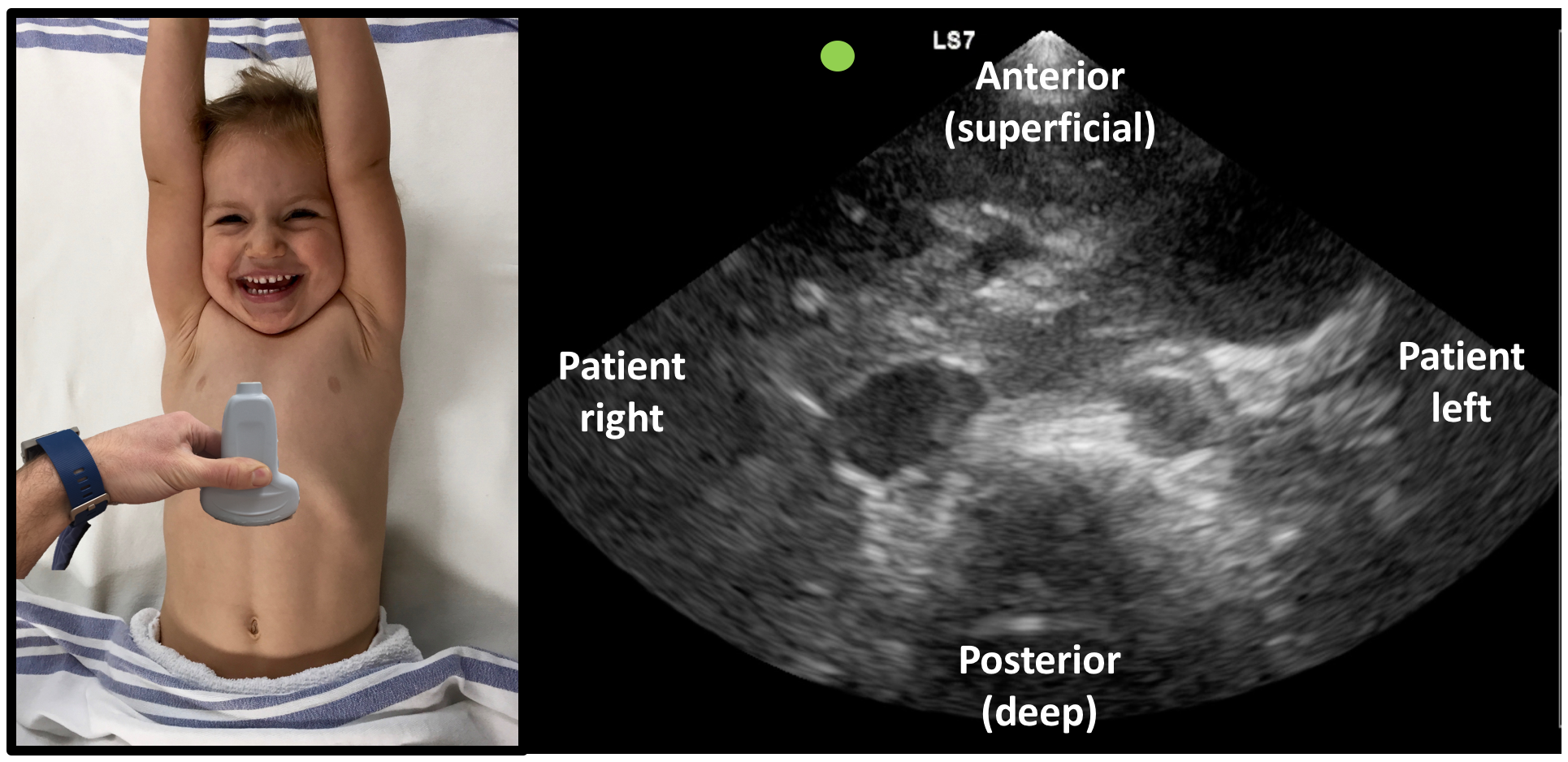
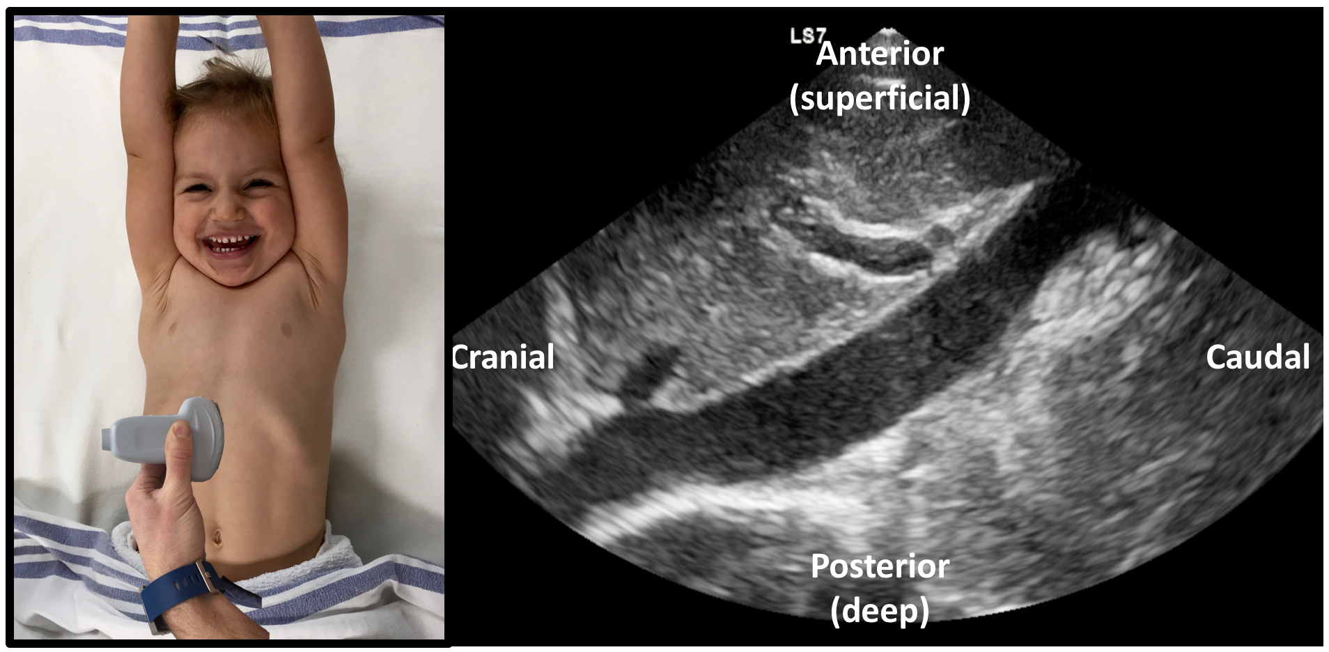
One of the tougher concepts to grasp is the orientation of the image on the screen in relation to the patient. Since the US probe can be moved and rotated to image patients in varying planes it is important to understand the relation of the image on the screen to the anatomy of the patient. In all cases the image on the top of the screen, or near-field relates to the superficial part of the patient that is touching the probe and the bottom of the screen, or far-field is deepest into the patient. Screen left corresponds to the direction the probe indicator is pointed on the patient, with the right screen being the opposite (Figure 13&14).
 Figure 13: Transverse view orientation
 Figure 14: Longitudinal view orientation
It is critical to have a proper reference to know how to interpret what we are seeing. One way to think of it is to imagine the probe is a flashlight looking into the body with the closest structures to the probe at the top of the screen and farthest at the bottom. You aim the flashlight towards the area of interest by manipulating your hand movements. Remind yourself where you are starting by mentally noting the corresponding structures to near-field, far-field, screen right and screen left. This cueing mechanism will become very useful once you start manipulating the probe to optimize images and scan through structures.
Optimizing images
The typical ultrasound machine found in North American hospitals will contain a bevy of buttons. These can be quite intimidating. Fortunately, for the majority of our indications we can focus on the few critical controls and ignore the rest. The most important controls to optimize the image are gain and depth.
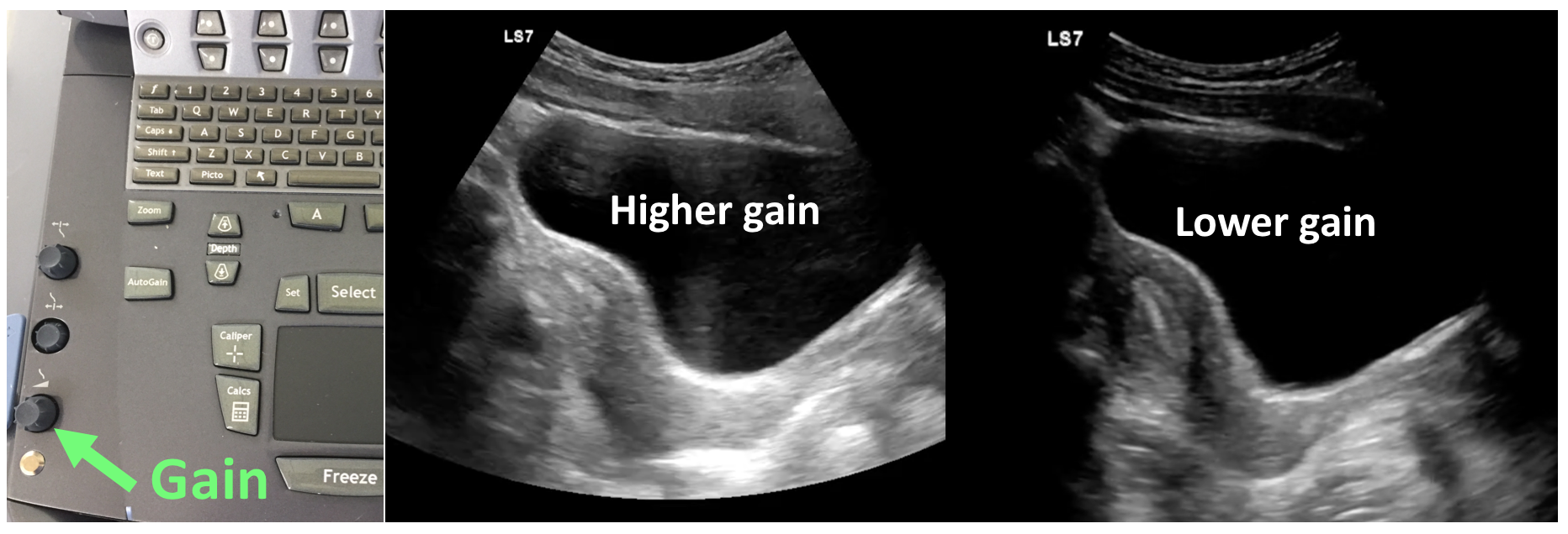
Gain controls how bright the image appears on the screen. Adjusting the gain essentially increases or decreases the sensitivity of the machine to ALL ultrasound waves sensed by the transducer. It acts as a “volume control” for the image brightness, turning up the gain makes everything appear brighter on the screen while turning it down makes everything darker.
 Figure 15: Gain
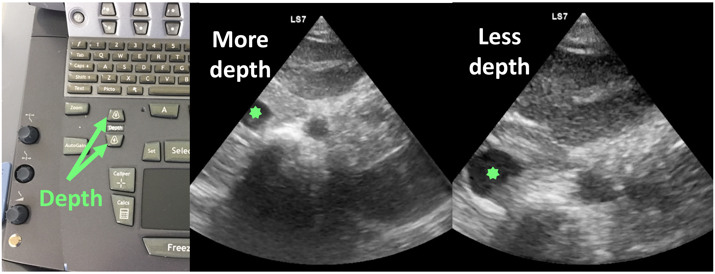
Depth controls how deep the ultrasound waves returning to the probe are detected. Increasing depth will allow you to see deeper into the patient, while decreasing help you focus more superficially (Figure 16). There is a numeric scale on the right side of the screen to help orient you to the depth of the image you are viewing. Often it is best to begin imaging an area of interest using the maximum depth and then gradually reduce the depth to optimize the visualization of the area of interest. This helps to landmark surrounding structures and avoid missing anything due to a superficial scan.
 Figure 16: Depth
