Evaluation of ETT depth
Evaluation of Lung Sliding
The depth of ETT insertion can also be evaluated using lung POCUS. Normal lung sliding indicates aeration of the lung. The movement caused by inflation of the lung can be seen as the visceral and parietal pleura move and is called lung sliding or shimmering. As such, presence of bilateral lung sliding indicates endotracheal intubation with an ETT positioned above the carina. The absence of lung sliding suggests the lung is not being ventilated and raises suspicion for esophageal intubation. If unilateral lung sliding is seen, it can suggest mainstem intubation [22].
Technique
Lung ultrasound can be performed with the patient in the supine position, using a linear or curvilinear probe placed in the longitudinal plane on the anterior chest of the patient at the midclavicular line. This is performed on both the left and right anterior chest walls. The relevant anatomic structures for this view are the pleural line, chest wall musculature and the ribs and their acoustic shadows (Video 3)
Important limitations to using lung sliding for confirmation of ETT depth include diseases and conditions that affect the pleura and therefore lung sliding on POCUS. This includes pneumothorax, ARDS, pneumonia, pleural disease and contralateral lung sliding despite mainstem intubation because of retrograde air movement.
Video 3. Lung ultrasound video performed using a linear probe placed in the longitudinal plan on the anterior chest of the patient at the midclavicular line demonstrating pleural line with normal sliding
Video 4. Lung ultrasound video performed using a linear probe placed in the longitudinal plane on the anterior chest of the patient at the midclavicular line demonstrating absent lung sliding
M-Mode can also be used to confirm the presence of lung sliding. In a normal lung where lung sliding is present, the “sand on the beach” or “seashore” sign can be observed (Figure 8A). Conversely, absence of lung sliding will create the “stratosphere” or “barcode” sign (8B).
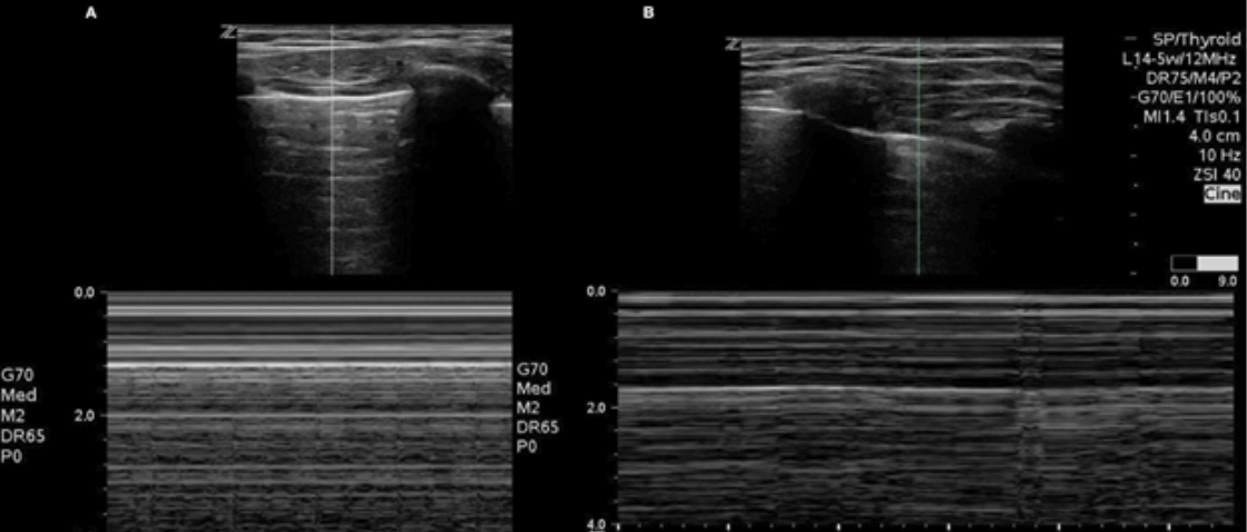
Figure 8. Use of M-mode to identify A) presence (seashore sign) or B) absence (stratosphere sign) of lung sliding
For more information on lung sliding and lung PoCUS, please revisit the KidSONO Pneumothorax Module.