What is NOT Normal?
Intussusception will appear differently due to the loss of air from the telescoped bowel. As a result, you are more easily able to visualize the intussusception compared to normal bowel. In the transverse view an intussusception appears as a “target” or “doughnut,” which is created by a hyperdense centre surrounded by multiple concentric hyperdense rings (Figure 6,8). This should have a minimum diameter of 2.5-3 cm with ileocolic intussusception. A “target” with a diameter of 2 cm or less suggests a small bowel intussusception, which generally self-resolve, and rarely require reduction. Intussusception appears as a “pseudokidney” or “sandwich” in the oblique or longitudinal view because of the multiple hypoechoic layers (Figure 10).
– The diameter of intussusceptions are typically estimated visually or “eye-balled.” However, if there is any uncertainty, callipers can be used to measure the anterior-posterior (AP) diameter from outer edge to outer edge in the transverse plane
In the case of a true ileocolic intussusception other findings can often be noted on ultrasound including small amounts of free fluid outside the intestine in the area of the intussusception as well as a small bowel obstruction. These findings are outside of the scope of this module and are discussed in the small bowel obstruction module.
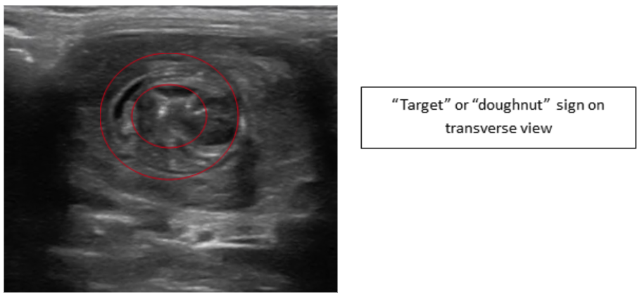 Figure 6: Transverse image intussusception—Target or Doughnut sign
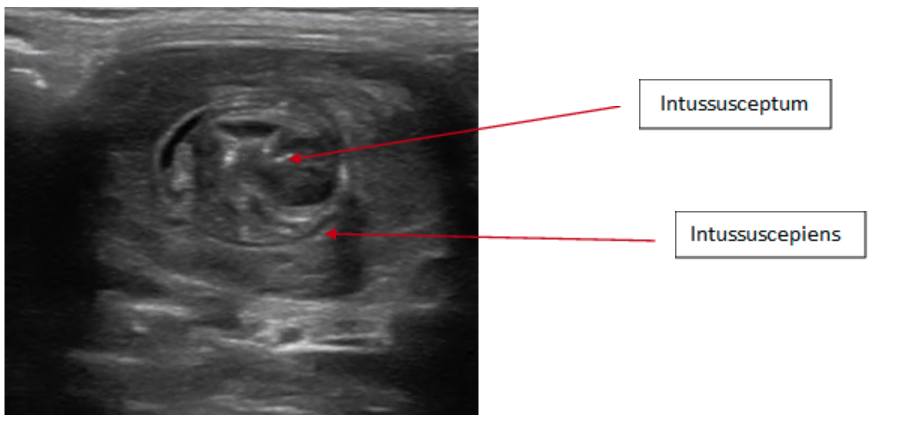 Figure 7: Transverse image intussusception, telescoping of the intussusceptum into the intussuscepiens
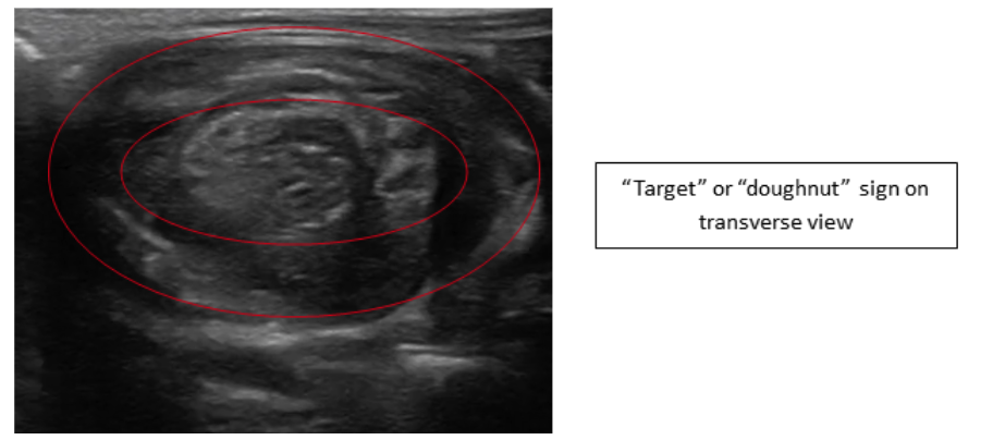 Figure 8: Transverse image intussusception—Target or Doughnut sign
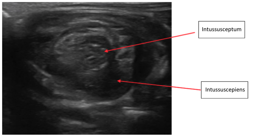 Figure 9: Transverse image intussusception, telescoping of the intussusceptum into the intussuscepiens
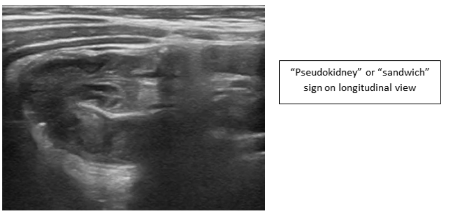 Figure 10: Intussusception “pseudokidney” or “sandwich” in the oblique or longitudinal view
Video 3: Normal bowel transitioning into Intussusception