What is NOT normal?
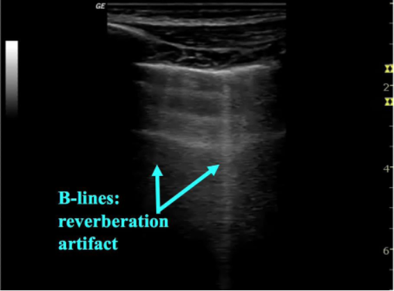
As pneumonia develops edema begins to collect in the lung, allowing for visualization of lung on ultrasound. B-lines represent thickening of the lung parenchyma—most often secondary to edema and appear as sharp, vertical lines arising from the pleura and extending to the base of the image on the screen. B-lines obscure A-lines and move in concert with lung sliding (figure 4). 1-2 B-lines per field of view can be normal in dependent regions or areas of interlobar fissures and should be clinically correlated. Localized B-lines may be the only finding early in disease course and they can often be found adjacent to areas of consolidation. While focal B-lines may indicate a bacterial pneumonia they are non-specific and can also be seen in viral etiologies. Additionally, when B-lines are found diffusely they are more likely to indicate other disease states such as cardiogenic pulmonary edema or viral pneumonia (8). B-lines and interstitial syndromes are discussed in detail in a dedicated KidSONO module.

Figure 4: B-lines, indicating interstitial thickening
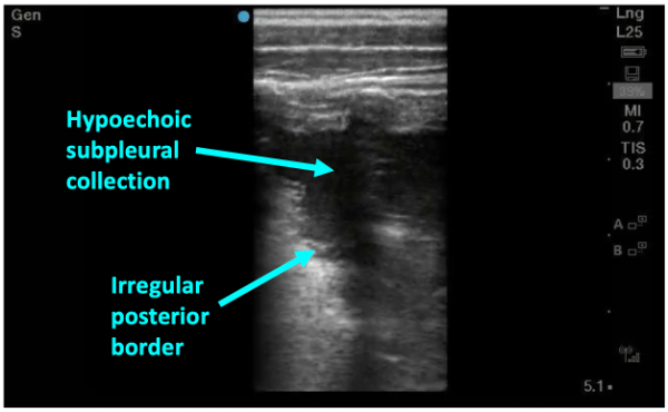
As pneumonia progresses and fluid starts to leak into alveoli, dark, irregular subpleural consolidations can be seen. They are backed by an irregular posterior border indicating transition from non-aerated to aerated alveoli—this is called the shred sign (figure 5). Subpleural consolidations that are greater than 1cm in diameter are generally considered to be indicative of bacterial pneumonia—smaller consolidations can also be found in viral processes such and bronchiolitis and viral pneumonias so clinical judgement is needed.

Figure 5: Sonographic consolidation with hypoechoic collection abutting the pleural line with an irregular posterior border– the “shred sign”
In more severe disease a dense area of consolidated lung begins to look like liver parenchyma on ultrasound. This is described as “hepatization” (figure 6).

Figure 6: Advanced disease with dense consolidation known as “hepatization” of the lung
Keep in mind LUS is also more sensitive than CXR for detecting pleural effusions, with just 10 mL detected on LUS, in comparison to 200mL required to be detected on CXR (9). If a parapneumonic effusion is present, LUS will easily find the anechoic area, typically at the base or more dependent areas. PoCUS for the detection of pleural effusions is covered in a dedicated module.
