Introduction
The most common initial test for respiratory distress is chest x-ray. Recently, growing evidence has shown that PoCUS can reliably detect thoracic pathology with equal if not better sensitivity than chest x-ray (CXR). In the case of pneumothorax PoCUS is significantly more sensitive than CXR. It also has the advantage of being performed at the bedside more quickly than CXR. Computed tomography offers little advantage over PoCUS, is impractical and comes with the cost of significant radiation.
Why Ultrasound?
Traditionally it was thought that ultrasound was not a useful modality for investigating lung pathology because air scatters ultrasound waves. There has been a growing body of evidence in recent years contradicting this practice. In fact, ultrasound images of normal lung show predictable artifacts that are disturbed in disease states and can be readily differentiated by ultrasound.
A recent meta-analysis comparing PoCUS to computed tomography in adults with pneumothorax found that in the hands of trained users PoCUS had a sensitivity of 91% and specificity of 98%. The same study found supine CXR had a sensitivity of only 50% [1]. While upright CXR may be more sensitive, it has never been formally compared to CT and the few studies available have found a sensitivity of 83-84% [2,3]. While these are mainly adult studies, PoCUS maintains its superior test characteristics in the neonatal population with novice sonographers who received one hour of training with sensitivities and specificities nearing 100% suggesting it is a useful tool throughout the lifespan [4,5,6].
Ultrasound has the additional benefits of being free from ionizing radiation and quicker to perform at the bedside. In a study of PoCUS versus CXR for neonatal pneumothorax ultrasound took on average 5 minutes versus 19 minutes with portable x-ray [4].
PoCUS is safe, timely and effective for the diagnosis of pneumothorax.
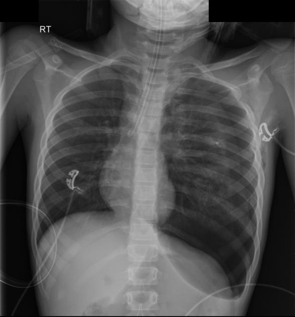 Supine CXR showing pneumothorax
PoCUS:
Sensitivity 91%
Specificity 98%
Supine CXR:
Sensitivity 50%
Specificity 99%
PoCUS vs CXR in trained users [1]