Parasternal Short Axis View
Technique
- The easiest way to find the PSAX view is to optimize your PSLX view and while maintaining the probe position on the chest wall rotate your probe 90 degrees clockwise (for cardiology convention) (Figure 6) or counterclockwise (for radiology convention) (Figure 7).
- The ultrasound probe should be placed immediately patient left of the sternum at at the level of the 3rd of 4th intercostal space
- To obtain a cross-sectional view of the heart the probe marker is directed to the right hip in emergency convention or the left shoulder in cardiology convention (resulting in the same image on the screen)
- Depth should be adjusted to ensure the posterior pericardium is visible in the far field
- Gain should be adjusted so that the myocardium appears grey, and blood appears black
- The probe should be adjusted using small movements of sliding, sweeping and rotation to optimize the view, ensuring the LV appears circular on screen and not oval or elongated.
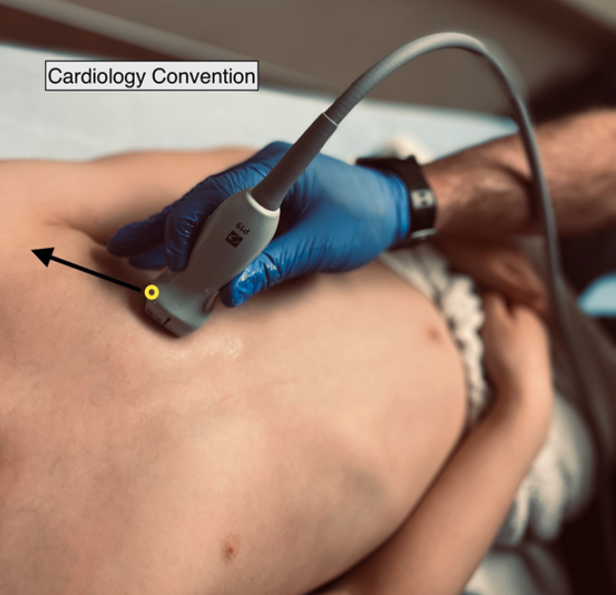
Figure 6. External Landmarking for parasternal short cardiology convention
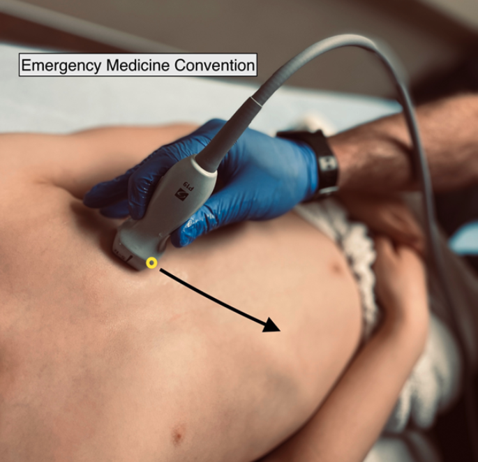
Figure 7. External Landmarking for parasternal short EM convention
Note: Emergency vs Cardiology convention:
The parasternal short axis view will appear the same on the screen whether you are using cardiology or emergency convention. This is because both the probe markers and screen markers are oriented opposite, resulting in the same net image on screen
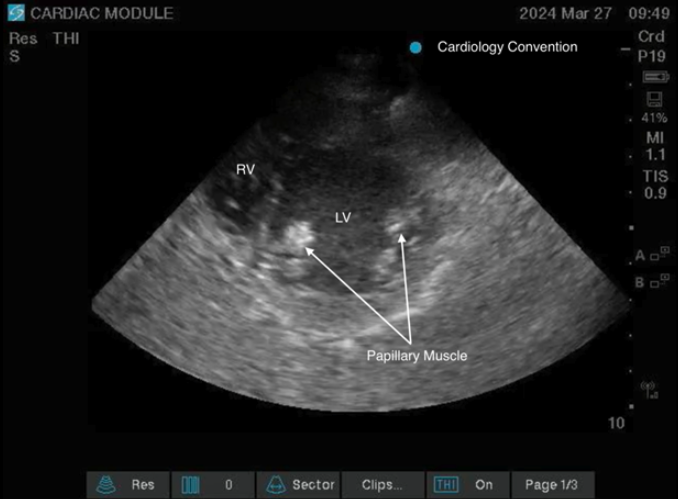
Figure 8: Parasternal Short View Cardiology Convention.
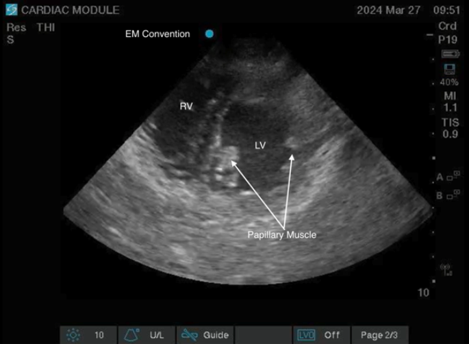
Figure 9: Parasternal Short View EM convention.
Scanning Tips:
- Sometimes it is necessary to slide the probe to different intercostal spaces to find the best window to view the heart, especially mechanically ventilated patients. If struggling, try each intercostal space starting from below the clavicle
- If struggling to find a window, lying the patient in the left lateral decubitus position will aid in pulling the heart against the chest wall and the lung away from it
- The easiest way to find the PSAX view is to optimize your PSLX view and while maintaining the probe position on the chest wall rotate your probe 90 degrees clockwise.
- Caution is required to avoid probe sliding when rotating the probe from the PLAX to obtain the PSAX view and to rotate 90 degrees to prevent an oblique view. Two hands can be used for this technique; one to rotate the probe while the other hand secures the position of the probe on the patient’s chest
What Am I Looking At?
In this view a cross section of the short axis of the heart is obtained. Most prominent is the circular LV, appearing as a donut in the middle of the screen. In the near field just below the probe the crescent-shaped right ventricle is visible, “hugging” the LV. The LV and RV are separated by the interventricular septum (Figure 10).
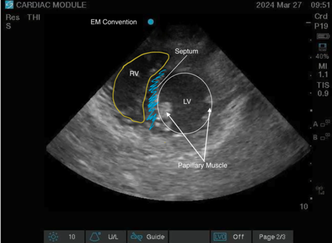
Figure 10: Parasternal Short View Labeled
PSAX Imaging Planes
Different imaging planes are available from the PSAX. Progressive sliding (angling if the window does not allow sliding) of the face of probe from the patient’s right shoulder toward the patient’s left hip will successively produce images from the base through the apex of the heart. Here we will focus on the fundamental images obtained from the PSAX, starting with structures most superiorly at the right shoulder and progressively lower to the left hip:
Aortic valve level: The aortic valve will be seen as a “Mercedes Benz” sign as the three leaflets of the AV close to produce the three-pointed star associated with the car logo. An ideal view will also include the RA, TV, RV, atrial septum and LA. (Figure 11)
Figure 11: Parasternal Short Aortic Valve Level
Mitral valve level: From the AV view, the ultrasound beam is tilted inferiorly until the mitral valve is visualized. The MV has been described as a “fish-mouth” with anterior and posterior mitral leaflets representing the upper and lower lip respectively (Figure 12). In this view, the RV, IVS, and LV are also visualized. (Figure 13)
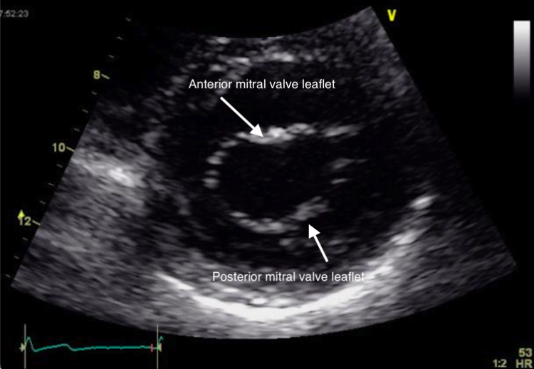
Figure 12: Parasternal Short Zoomed Mitral Valve
Figure 13: Parasternal Short Mitral Valve View Video with color in the cardiology convention
Mid-papillary level: Further tilting of the ultrasound beam in an inferior direction will produce the mid-papillary image. The papillary muscles appear as slightly hyperechoic “knobs” protruding from the inferior aspect of the LV. The posteromedial papillary muscle (PM) position will vary between the 6-9 o’clock positions, while the anterolateral PM is usually seen between the 3-6 o’clock position. The RV, IVS, and LV are also visualized. (Figure 14)
- In the critically ill patient, the mid-papillary view has the highest yield and provides the best assessment of global LV function and any wall motion abnormalities. This is the gold standard view for evaluating the shape of the interventricular septum.
Figure 14: Parasternal Short Mid-Papillary View
Apex: Finally, further tilting the ultrasound probe inferiorly until the PMs are no longer seen will generate a view of the LV apex. The RV and IVS are also visualized. (Figure 15)
Figure 15: Parasternal Short Apex View
Clinical Utility
The PSAX view at the mid-papillary level provides global assessment of LV function and any LV wall abnormality. The mid-papillary view also allows visualization and assessment of the interventricular septal shape when assessing the right side of the heart.
