Anatomy Review: What am I looking at?
The appendix is a small, blind-ended tubular structure. The appendix typically arises from the posteromedial aspect of the cecum, just below the ileocecal valve. The tip can have variable position within the right lower quadrant and is often found below or slightly posterior to the terminal ileum and anterior to the iliac vessels.
Less commonly, the appendix can be retrocecal, extending behind the cecum making visualization more difficult, or have a pelvic position, where it extends downward into the pelvis, closer to the bladder and reproductive organs. Additionally, it can sometimes extend laterally from the cecum, which may also affect its visualization.
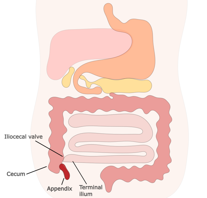
Figure 1: Typical anatomical position of the appendix in the RLQ
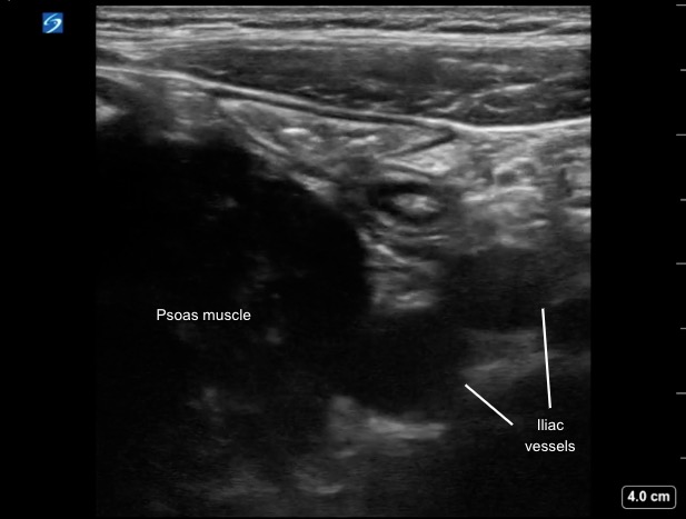
Figure 2: Transverse image of the RLQ demonstrating anatomical landmarks (Iliac vessels & Psoas muscle) used to identify the appendix on PoCUS
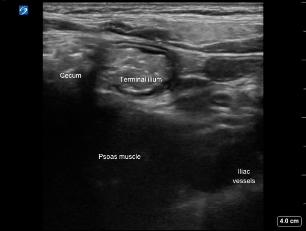
Figure 3: TTransverse image of the RLQ anatomy, showing the cecum positioned lateral to the psoas muscle and the TI medially—key landmarks for locating the appendix on PoCUS
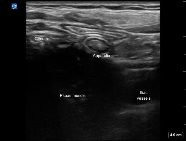
Figure 4: Transverse image of the RLQ showing the “typical” position of the appendix
Figure 5: Transverse video of the RLQ anatomy