Testicular Anatomy Review
In a patient with normal anatomy (Figure 1A), the scrotum is divided into left and right by the scrotal septum. Each compartment contains a testis, an epididymis, and the spermatic cord. The testicle is covered by a fibrous capsule called the tunica albuginea. This capsule projects into the testicle to form the mediastinum testis. Testicular seminiferous tubules run through the mediastinum testis and exit the testicle to form the epididymis which continues as the vas deferens. The spermatic cord contains the vas deferens as well as testicular vessels and nerves.
In testicular torsion, the testicle spins, twisting the spermatic cord and causing compression of the blood vessels (Figure 1B), thereby limiting venous outflow and arterial inflow (Figure 1C).
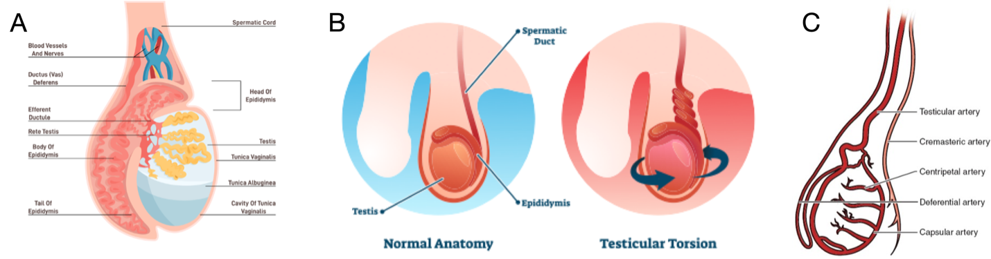
Figure 1. A) Normal testicular anatomy. B) Testicular torsion demonstrating torsion of the spermatic cord C) Arterial supply to the testicle