
Patient Position: Lying down or sitting with parent
Patient preparation:
- Consider swaddling an infant or burrito wrapping a toddler
- Have the parent hold the child’s head or distract the child in to facilitate scanning the area of interest
Stepwise Technique:
1. Position the transducer over the area of swelling/tenderness
· While the transducer can technically be orientated in any direction, consistency is important for interpretation and documentation
· When possible, start with aligning the probe marker toward the patient’s right
2. Identify the skull bone
· Confirm the skull line appears as a crisp, hyperechoic line
3. Scan from normal (no swelling) through abnormal and back to normal
· *Ensure to scan 2cm beyond borders of swelling as a fracture may be located outside the area of swelling
4. Rotate and repeat the process in a perpendicular plane.
· Rotate the probe 90 degrees from the starting position and perform the same assessment in the perpendicular plane.
· The probe can be rotated 90 degrees in either direction—choose the direction that best suits the scanning position and your ability to control probe movements.
· When possible, aligning the probe marker toward the patient’s face
** Clearly indicate probe orientation and position on images to avoid confusion
5. Document and review images/clips for both immediate assessment and for review afterwards.
· Findings can be subtle so view and record while scanning but ensure to review images after. If in doubt scan the area further
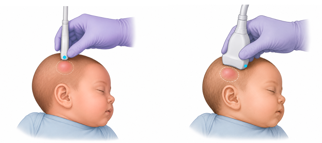 Figure 2ab. Probe orientation. Starting position with the probe marker oriented toward the child’s right (a), followed by a 90° rotation (b).