The subxiphoid 4 chamber view is particularly well suited for RV assessment due to the anterior position of the RV and the ability to use the liver as an acoustic window. This view provides excellent visualization of the RV free wall and is especially useful for evaluating RV free wall motion, wall thickness, RV size and global function.
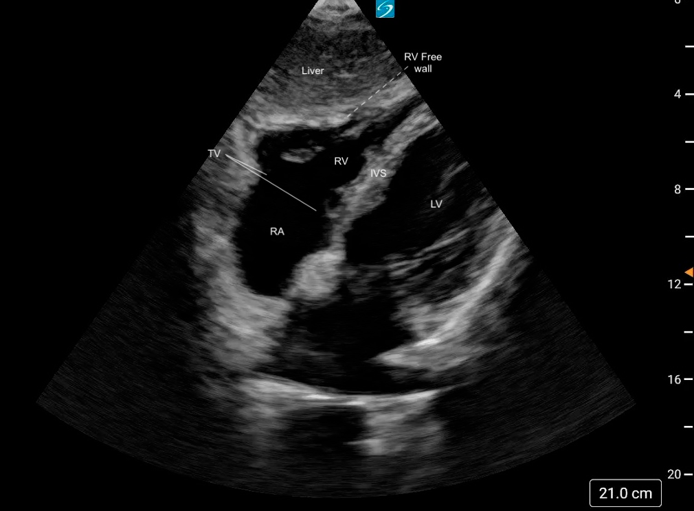 Figure 17. Normal subxiphoid 4 chamber view labeled
What is Normal?
- Triangular shape
- Thin walled
- Smaller than the LV: A general rule of thumb is that the RV should be ~2/3 the size of the LV [19].
- The RV should squeeze inwards uniformly, with the free wall moving toward the septum during systole.
Figure 18: Subxiphoid 4 chamber view demonstrating normal RV size and function
What is NOT Normal
- Visually, if the RV is equal to or larger than the LV, then there is likely RV dilation.
- Visually reduced contraction.
- Decreased or akinetic RV free wall motion (Mcconnell’s Sign)
Figure 19: Subxiphoid 4 chamber view demonstrating dilated RV with reduced function