What is Normal?
The normal pediatric eye appears symmetric and well-structured, with clear chambers, a centered lens, and an intact retina on ultrasound. Because a generous amount of gel is used in ocular ultrasound to minimize pressure on the eye, a thin layer of gel will be visible on the image. The eye will have the same appearance in both sagittal and transverse planes, reflecting its round, symmetric shape.
- Eyelid: Thin, echogenic superficial layer.
- Anterior Chamber: Anechoic, uniform depth, no internal echoes.
- Iris: Thin echogenic line anterior to the lens.
- Posterior Chamber: Anechoic space immediately posterior to the iris.
- Lens: Bright, round, echogenic posterior border with an anechoic center. Symmetric and centered behind the iris.
- Vitreous: Uniformly anechoic, without internal echoes.
- Retina: Thin echogenic line along the posterior globe; flat and attached.
- Optic Nerve: Hypoechoic tubular structure extending posteriorly from the globe.
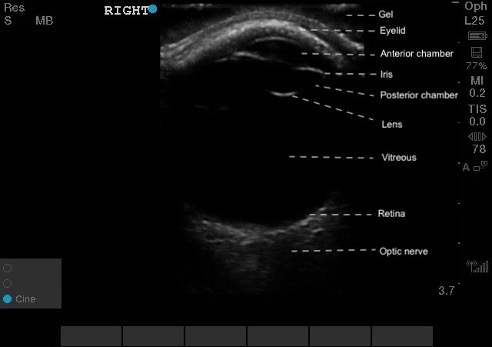 Figure 5. Normal ocular ultrasound of the right eye, labeled.
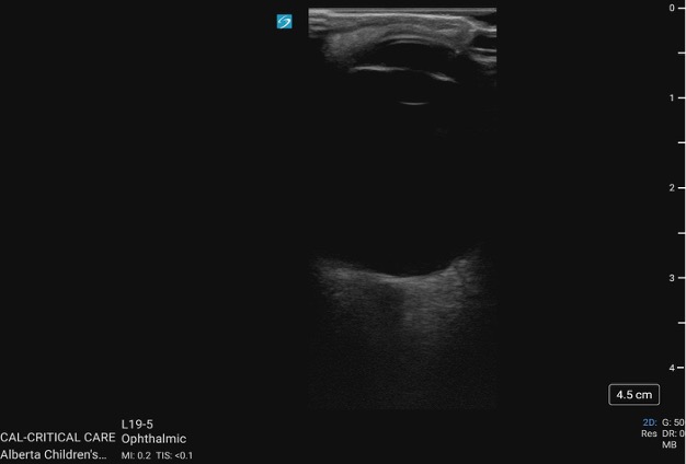 Figure 6. Normal eye on PoCUS.
Ocular Movements (figure 7):
Eye movements are seen as smooth, coordinated shifts of the globe. The lens and iris move together, and the retina remains attached and stable. Uniform motion in all directions indicates normal extraocular muscle function.

Figure 7. Normal ocular movements seen on ultrasound