Hydronephrosis
Hydronephrosis is defined as an abnormal enlargement and distension of a kidney caused by obstructive uropathy (blockage of normal urine flow into the bladder). Patients may present with varying symptoms depending on the causes of hydronephrosis [10]. On ultrasound, this appears as abnormal distension of the renal pelvis which is normally not visible within the fat of the renal sinus. Depending on the degree of obstruction, hydronephrosis can be graded according to severity.

Grade 1 Hydronephrosis or “Mild” Hydronephrosis
Occurs when there is dilatation of the renal pelvis (pelviectasis) without dilatation of the calyces. The renal cortex (parenchyma) is preserved and does not show any atrophy.
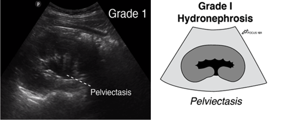 Figure 11: Longitudinal ultrasound image of the kidney with corresponding anatomical diagram, demonstrating pelviectasis. Image use with permission POCUS 101 [10].
Grade 2 Hydronephrosis (Mild-Moderate)
Occurs when you have dilatation of the renal pelvis (pelviectasis) and dilatation of the calyces (caliectasis), specifically the MAJOR calyces. The renal cortex (parenchyma) is preserved and does not show any atrophy.
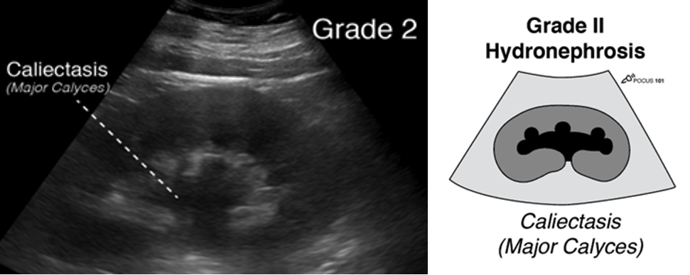 Figure 12: Longitudinal ultrasound image of the kidney with matching anatomical illustration, showing pelviectasis and dilation of the major calyces. Image use with permission POCUS 101 [10].
Grade 3 Hydronephrosis or “Moderate” Hydronephrosis
Occurs when you have dilatation of the renal pelvis (pelviectasis) and dilatation of the calcyces (caliectasis), specifically the major and minor Mild cortical thinning may be seen.
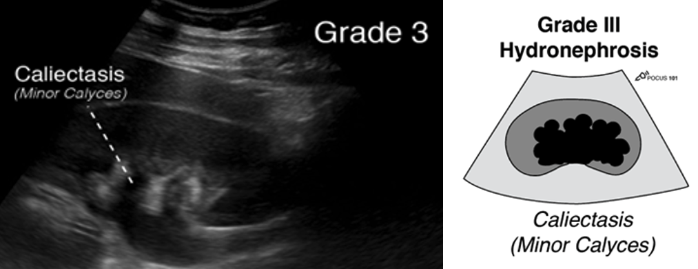 Figure 13: Longitudinal ultrasound image of the kidney with matching anatomical illustration, showing pelviectasis and dilation of the major and minor calyces. Image use with permission POCUS 101 [10].
Grade 4 Hydronephrosis or “Severe” Hydronephrosis
Occurs when there is significant/gross dilatation of the renal pelvis (pelviectasis) and calcyces (caliectasis) resulting in renal cortical thinning. There will also be renal atrophy and loss of the borders between the renal pelvis and calyces.
· Grade 4 hydronephrosis is also sometimes called the “Bear Claw” sign since the anechoic areas resemble a paw print.
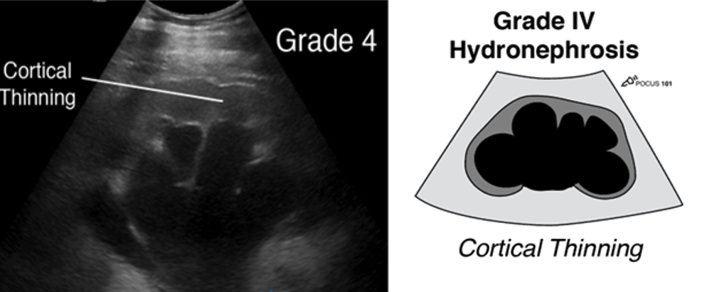 Figure 14: Longitudinal ultrasound image of the kidney with matching anatomical illustration, showing pelviectasis, caliectasis and cortical thinning. Image use with permission POCUS 101 [10]
Absence of Ureteral Jets
In cases of obstructive uropathy, either from stones or other causes, absence of ureteric jets in addition to hydronephrosis can be appreciated in the bladder indicating a blockage urine passing from the kidney to the bladder. Absence of jets is highly suspicious for obstructive uropathy and formal imaging follow up with radiological US or CT scan is recommended.
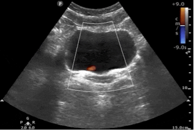 Figure 15: Transverse bladder with absent left uretic jet
Ultrasound Findings for Kidney Stones
PoCUS for the direct visualization of kidney stones is limited. Formal radiology has high sensitivity and specificity for identification of stones depending on the experience of the institution but is generally not reliable at the bedside. Sometimes renal stones can be seen in the renal cortex, pelvis, or the ureteropelvic junction appearing as a hyperechoic structure with posterior shadowing.
The “twinkling artifact” is a sonographic artifact located behind calcifications of ureteral calculi when color Doppler is applied. It will appear as a multicolored high-intensity signal, like signals produced by turbulent flow with aliasing. The twinkling artifact has a sensitivity and specificity of 90% and 100%, respectively, for kidney stone at the ureterovesical junction [14]. The twinkling artifact should not be confused with ureteric jets, which are found within the fluid of the bladder, not behind the posterior wall.
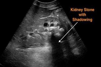 Figure 16: Sagittal image of the right kidney with a large stone displaying posterior enhancement in the lower pole. Image use with permission POCUS 101 [10].
 Figure 17: Transverse color image of the bladder showing twinkle artefact at the left UVJ indicative of a stone. Image use with permission from POCUS Atlas [12]