Left Kidney
Technique
The technique for scanning the left kidney is very similar to that of the right kidney. Of note, the left kidney lies slightly superior, and posterior compared to the right kidney due to the smaller size of the spleen.
- Identify the kidney in the longitudinal view:
· Place the probe along the left posterior axillary line around the level of the xiphoid with the probe indicator directed towards the patient’s head
· Slide the probe until the spleen comes into view: the left kidney is located more superior, and posterior compared to the right kidney. To view the left kidney, the scanner’s knuckles are often touching the bed in the posterior axillary line
· Once the spleen is visualized slide, fan and heel the probe to identify the left kidney and center it on the screen
· Adjust depth and gain if necessary and magnify the kidney to optimize the image

- Assess the LK in transverse view:
· From sagittal, rotate the probe 90 degrees counterclockwise so the probe marker is oriented anteriorly
· Slide or fan the probe from cranial to caudal to assess the kidney in its entire transverse view
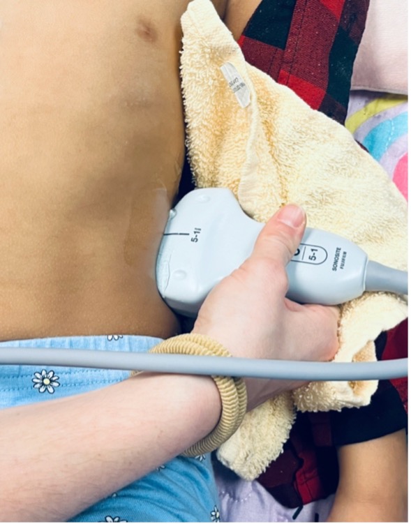
Figure 5: Patient/probe position for sagittal assessment of the left kidney
What am I Looking at?
The left kidney is located just inferior and slightly medial to the spleen. The spleen appears homogeneously echogenic with a uniform texture and is less vascular than the liver. On ultrasound, the spleen is typically slightly more echogenic than the left kidney.
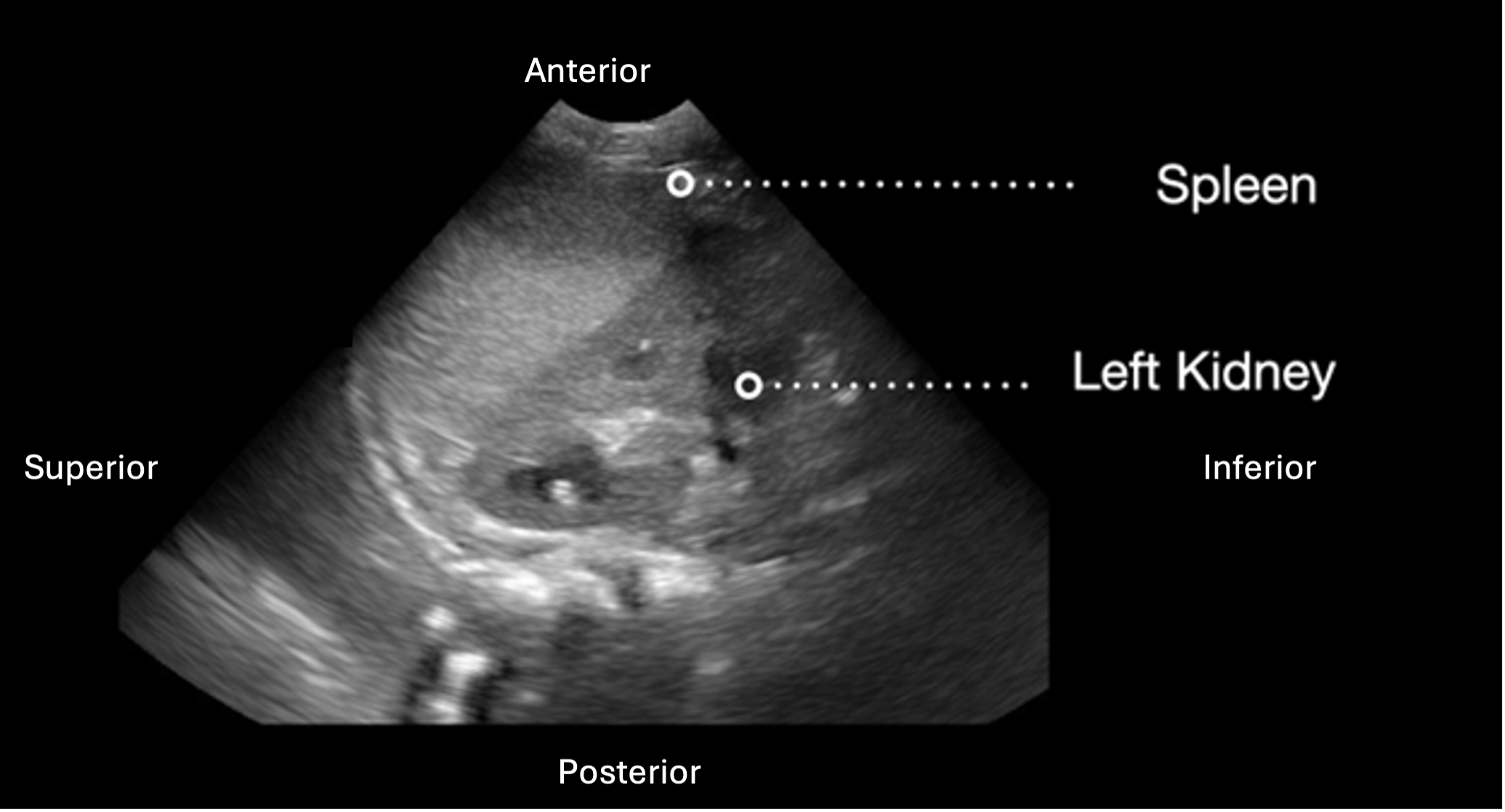 Figure 6: Longitudinal view of the left kidney with labeled image orientation and surrounding structures
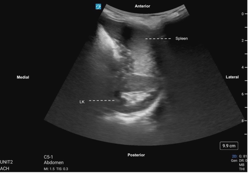 Figure 7. Left kidney transverse, labeled with image orientation and surrounding structures
Figure 8: Transverse sweep of the LK