Right Kidney
Technique
- Identify the kidney in the longitudinal view:
· Place the probe in the right midaxillary line at the level of the xyphoid process with the probe indicator towards the patient’s head.
· Slide the probe posteriorly and anteriorly until the kidney is located
· Adjust the view by sliding the probe to ensure the kidney is centered in the field of view.
· Adjust depth and gain if necessary and magnify the kidney to optimize the image.
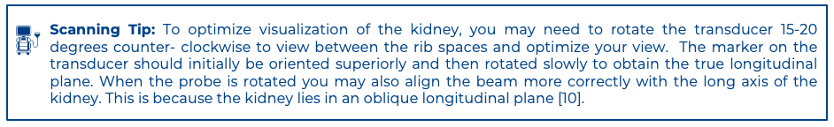
- Assess the RK in transverse view:
· From sagittal, rotate the probe 90 degrees counterclockwise so the probe marker is oriented posteriorly
· Slide or fan the probe from cranial to caudal to assess the kidney in its entire transverse view
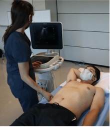
Figure 1. Patient and probe position for right kidney assessment. Image use with permission POCUS 101 [10].
What am I Looking at?
The right kidney is located just inferior and slightly lateral to the liver. The liver appears homogeneously echogenic with a smooth, uniform texture and contains prominent vascular structures. The right kidney is slightly less echogenic than the liver
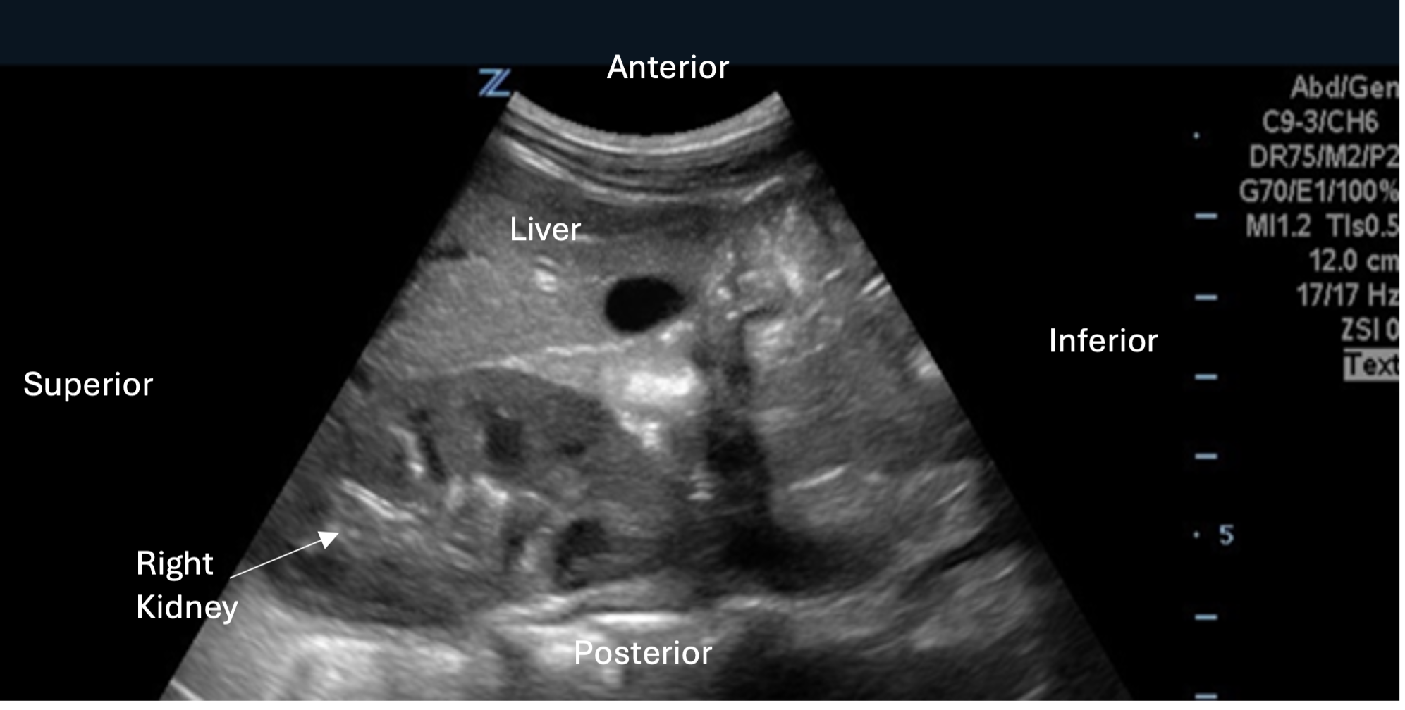 Figure 2. Longitudinal view of the right kidney with labeled image orientation and surrounding structures
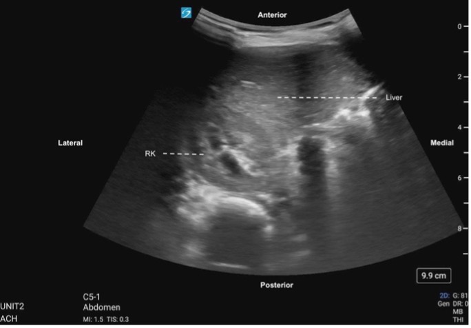 Figure 3. Right kidney transverse, labeled with image orientation and surrounding structures
Figure 4: Right kidney transverse