What is normal?
The normal lung is air-filled. Because air scatters ultrasound waves, we use the presence of normal artefacts to assess the lung. Depending on the window used, the lung’s appearance may vary. When viewed directly, the lung refracts US waves, but reverberation artefact creates A lines and occasionally B-lines (figure 5).
A-lines (figure 4):
- Reverberation artefact
- Ultrasound waves bounce back and forth between the probe and reflective pleura
- Results in regularly spaced, horizontal hyperechoic lines deep to the pleural interface
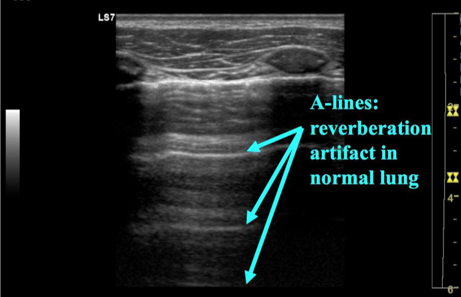 Figure 4: A-lines
B-lines (figure 5):
- A focal reverberation phenomenon
- Ultrasound wave bounce between the pleura and pulmonary interstitium
- Results in vertical lines from pleura towards the far screen that move with respiration
- Not always seen and are pathologic when numerous.
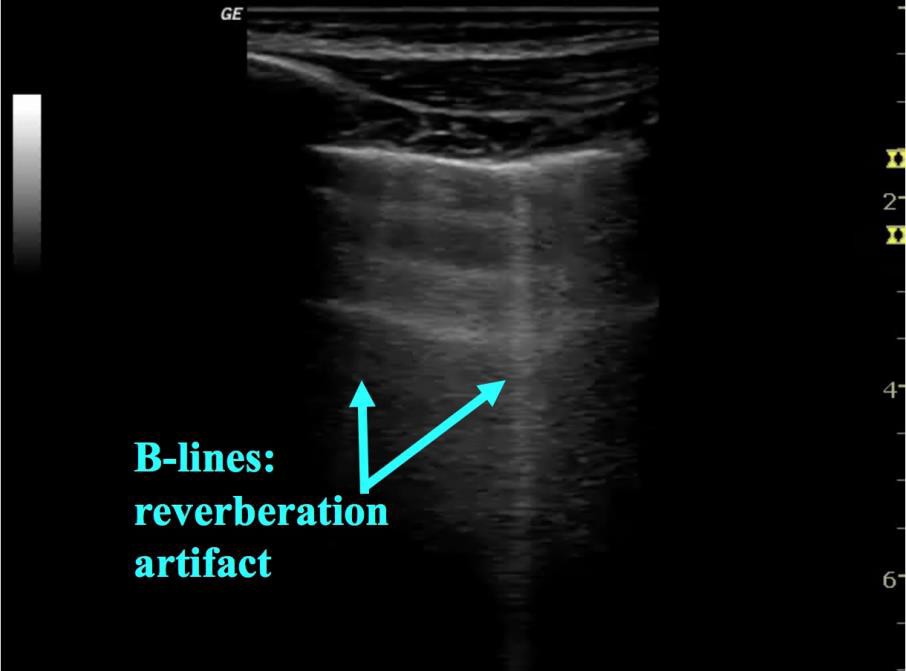 Figure 5: B lines
When viewed via the abdomen ultrasound waves are reflected by the diaphragm and create a mirror image effect: the lung appears as a liver-like organ above the diaphragm (figure 6). This artifact moves with respiration as the diaphragm moves up and down.
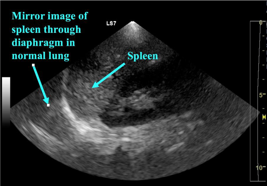 Figure 6: Mirror Artifact
In the absence of pathology, aerated lung fills the costophrenic angle and obscures the diaphragm in the nearfield. As the diaphragm descends with respiration, the air-filled lung crosses the screen and further obscures the diaphragm and structures behind. This is referred to as the curtain sign (video 2).
Video 2: Curtain sign