Apical Four Chamber View
Technique
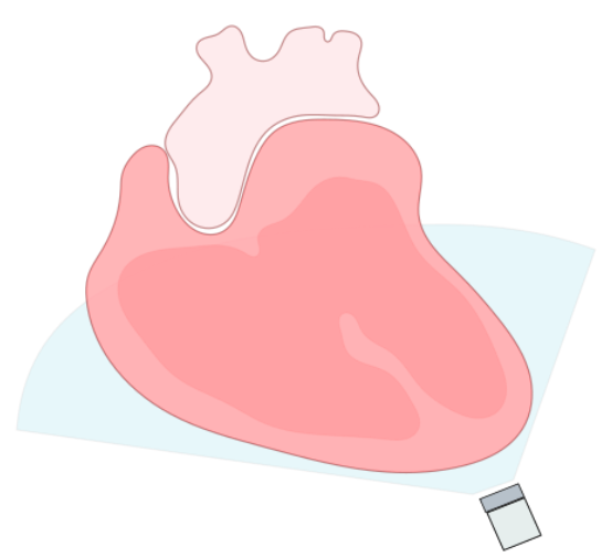
- In this view the heart is imaged in a coronal plane (Figure 16)
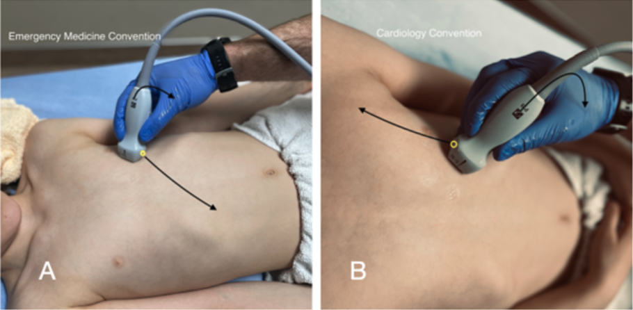
- To obtain an apical four-chamber (A4C) view, the probe is placed over the apex of the heart which is usually located in the vicinity of the left nipple (or inframammary line in females), in the 4th-5th intercostal space and the ultrasound beam directed towards the patient’s right shoulder.
- In emergency convention the probe marker is pointing towards the patient’s right hip and in cardiology convention it is directed towards the left shoulder (Figure 17)
- Once you reach the apex of the heart, as indicated by the left ventricle decreasing in size, tilt the tail of the probe down towards the patient’s feet.
- Depth should be adjusted to ensure visualization of the atria in the far field
- Gain should be adjusted so the myocardium appears grey and blood black.
- The ideal view should have the IVS centered on the screen and from the top to bottom on the screen, parallel to the ultrasound beam.

Figure 16: Coronal view of the heart from the apical window
Note: Emergency vs Cardiology convention
The apical four chamber view will appear the same on the screen whether you are using emergency or cardiology convention. This is because both the probe markers and screen markers are oriented opposite, resulting in the same net image on screen

Figure 17. External Landmarking for A4C View Emergency Medicine Convention (A) vs Cardiology Convention (B)
Scanning Tips:
- This window can be challenging to obtain, especially in mechanically ventilated patients, use lots of gel and make small circular movements until the best window is obtained.
- If struggling to find a window, lying the patient in the left lateral decubitus position will aid in pulling the heart against the chest wall and the lung away from it
- An adequate view should have the apex in the near-field, the ventricles appearing elongated with a straight interventricular septum running vertically down the screen.
- The position of the cardiac apex is highly variable. One method used to reliably obtain an adequate A4C is to begin with a high-quality PSAX and slide the transducer inferolaterally keeping the LV centered on the screen before tilting the face of the probe upwards to the right shoulder to view the heart in a coronal plane
What Am I Looking At
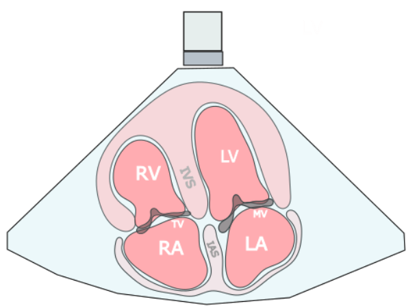
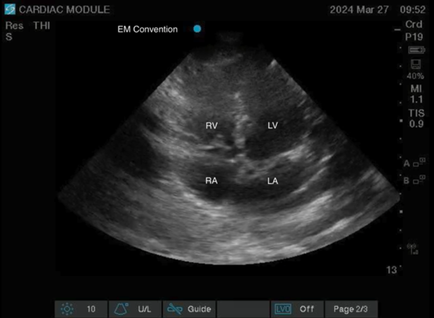
In this view a coronal view of the heart is obtained. In the near field just below the probe the apex with the RV visualized below on screen left and the LV on screen right. Deep to the ventricles the mitral and tricuspid valves can be separating the ventricles from the atria which lie in the far-field. The intraventricular and atrial septums can be seen running vertically on the screen from the near to far field, dividing the right and left sides of the heart.

Figure 18: A4C coronal view of the heart, illustrating the right and left atria (RA, LA), right and left ventricles (RV, LV), interventricular septum (IVS), interatrial septum (IAS), mitral valve (MV), and tricuspid valve (TV).

Figure 19: Apical Four Chamber View EM Convention
Figure 20. Apical Four Chamber View Video Clip
Clinical Utility
The apical four chamber view provides a wealth of information including global assessment of LV and RV function and size. It is the best view to compare ventricular sizes. This is another view in which pericardial effusions can be seen. The A4C view also allows two-dimensional evaluation of the tricuspid and mitral valves.
