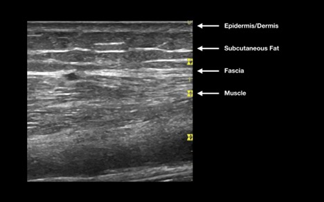
What is NOT normal?

What is NOT normal?

Cellulitis

Cellulitis can have a highly variable appearance. In general, the subcutaneous tissue appears thickened and will have a “cobblestone” appearance. This is caused by subcutaneous edema and inflammation generated by the surrounding infection. Cobblestoning is not a specific finding to cellulitis but a finding in tissue edema, care must be taken to correlate to the clinical scenario. If uncertain, compare the area of interest to an unaffected area of skin.


Figure 3: Cobblestoning

Cobblestone appearance of cellulitis Cobblestone sidewalk


Video 4: Soft tissue cobblestoning


Pitfalls

Edema from any source including:

  • Insect bites
  • Trauma
  • Generalized edema due to systemic causes

Abscess

Abscesses appear as heterogeneous anechoic, isoechoic (gray) or hypoechoic fluid collections. Their appearance is often not uniform. Abscesses can be well-circumscribed or be irregularly shaped areas of fluid which may demonstrate posterior acoustic enhancement (bright white in the far field). As with cellulitis, measure the size of the abscess in both the transverse and longitudinal plane. There is some evidence from an immunocompetent pediatric emergency department population that abscesses larger than 5 cm in diameter predict need for hospitalization [14]. In addition, measure the depth from the skin and the width from the base in order to guide future attempts at incision and drainage. As part of the evaluation, apply firm and graded pressure with the probe over the abscess to look for fluctuance (often over a point of maximal tenderness) and the “swirl sign.” Swirl sign refers to the movement of fluid and debris within the abscess and is generated by compression over the area. Finally, always evaluate the abscess with color Doppler to look for internal blood flow and surrounding vasculature prior to making an incision.


Video set 5: Examples of soft tissue abscesses

Courtesy of EDSONOshare library


Video 6: Video illustrating a positive “Swirl Sign”


Practice Pearl: Prior to performing any incision and drainage of an abscess, use color Doppler to look for evidence of blood flow. In addition, color Doppler can be used to identify surrounding blood vessels in order to decrease the rate of procedural complications.


Figure 4: Color Doppler over a suspected abscess

This mass was a clinically suspected abscess but bedside PoCUS identified significant blood flow. Planned incision was aborted and the patient was referred to surgery for further management.

Pitfalls:

  • Hematoma
  • AV malformation
  • Lymph nodes
  • Cysts

Necrotizing Fasciitis

Due to the low incidence and high morbidity of necrotizing soft-tissue infections, it remains a diagnosis that requires physicians to maintain a high clinical index of suspicion. The use of bedside ultrasound should never delay surgical consultation for these patients but should instead be used to expedite diagnosis. There are three main sonographic findings in necrotizing fasciitis. These include subcutaneous air (often tracking along a fascial plane) with accompanying “comet-tail” artifact, diffuse subcutaneous tissue thickening (which often requires comparison to a contralateral side), and >5mm of fluid accumulation along a facial plane [10].


Figure 5: Sonographic findings suggestive of necrotizing infection


Video 7: PoCUS suggesting necrotizing infection

Courtesy of EDSONOshare Library


Foreign bodies

Research has shown that over one-third of hand foreign bodies are missed at the index visit. Bedside ultrasound can be used to both diagnose foreign bodies and plan removal by accurately identifying the depth of the target and surrounding structures which may need to be avoided (i.e., vasculature). Ultrasound is superior to plain films for radiolucent objects such as plastic, wood or plant matter. When using ultrasound to guide removal of a foreign body, be sure to measure the distance from the skin surface with the calipers and scan the object in 2 planes to fully characterize its length, depth and width.

Practice Pearl: When using ultrasound to guide removal of a foreign body, consider placing multiple high gauge needles to mark the location of the object prior to dissecting down to find and then remove the foreign body. This can save a lot of time and avoid unnecessarily damaging tissue.


Figure 6: Foreign bodies on bedside ultrasound

Wood Glass Metal
Wood often demonstrates attenuation artifact or acoustic shadow. Glass often demonstrates reverberation or comet tail artifact. Metal often demonstrates reverberation artifact on ultrasound.


Video set 8: Soft tissue foreign bodies

Wood

Glass

Metal

Courtesy of EDSONOshare library


Pitfalls:

  • Fascia
  • Bones
  • Subcutaneous air